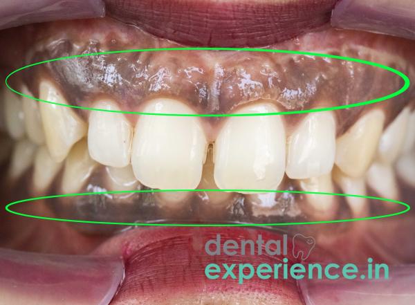
The discoloration of gums due to increased melanin production causes black gums. Healthy gums will typically be a shade ranging from light pink to coral. An increase in melanin production in the basal layer of the oral epithelium results in the gums turning a brownish-purplish-black. In periodontology, it is generally considered to be a normal variation as opposed to being abnormal; however, rapid or unexplained changes could suggest a variety of systemic or local issues such as toxicities in the body or localized trauma.
Definition: Darkening of the gums due to increased production of melanin by melanocytes in the basal layer of the oral epithelium.
Warning Signs: Sudden development of dark areas on the gums, bleeding upon gentle toothbrush application, redness, pain, or swelling around the area of darkened tissue, lack of healing after injury to the mucous membrane of the mouth.
Treatment: Depigmentation with lasers, surgical excision of the affected area by scalpel or electrosurgical knife and elimination of contributing factors.
Individuals with gingival pigmentation often first note obvious visual and tactile alterations:
Visual Changes: Evenly distributed throughout the attached gingiva or uneven, blotched dark brown or black lesions.
Changes in Texture: Uniform smooth surfaces, or irregular textures that have become inflamed and lost their stippling (natural stippling provides a healthy orange peel type texture).
Increased Vascularity: Darkened tissues that bleed easily upon routine brushing especially when they develop localized plaque buildup.
Dark gums resulting from benign ethnic pigmentation is generally not harmful. Seek emergency consultation with a periodontist if you experience:
Unilateral Growth: Quickly developing dark colored areas that vary in size and shape, have raised edges, or spread asymmetrically (potential indicator of oral melanoma).
Additional Symptoms: Halitosis (bad breath) or purulent discharge (pus) along with darkened tissue that bleeds during routine brushing, loose teeth, etc.
Systemic Indicators: Along with dark pigmentation of the buccal mucosa and/or tongue, chronic fatigue, and/or weight loss (possible signs of endocrine disorder such as Addison's disease).
The causes of gingival hyperpigmentation include physiological (basal melanin production), habitual/behavioral (tobacco use), and environmental (cultural/lifestyle factors) influences:
Basal Melanin Production: Physiologic stimulation of melanocytes (cells producing melanin) in the basal layer of the gingival tissue.
Nicotine-Stimulated Hyperpigmentation: Nicotine stimulates melanocytes to generate excess melanin as a protective response; tar from cigarettes stains oral tissue directly.
Amalgam Tattoos: Accidental placement of dental amalgam into the soft tissue during previous fillings creates permanent blue/black focal points.
Culture/Lifestyle Factors: Repeated chewing of paan (betel leaf) or smokeless tobacco products can cause chronic irritation to the mucosal lining and stimulate reactive melanization.
Medications: Certain long-term pharmacological agents — e.g., antimalarial drugs, minocycline, or certain psychotherapeutic agents — can induce this condition.
Tobacco use (both smoking and chewing): Tobacco use accelerates melanocyte function greatly.
Genetic Influence: Individuals of South Asian/Middle Eastern/African descent tend to have higher baseline melanin concentrations in their gums.
Chronic Gingivitis: Localized inflammation leads to elevated blood flow through the affected tissue and increased oxidative stress leading to enhanced melanin deposition.
Hormonal Fluctuations: During pregnancy and other periods of hormonal fluctuation there is a system-wide shift in the rate of melanin production.
Warning: Don't attempt to remove dark gum tissue by scratching/scraping/bleaching. This can lead to serious damage to your gum tissue.
Cleanse: Thoroughly wash your hands with antibacterial soap.
Reveal: Using clean fingers gently pull up your top lip and down your bottom lip to show your entire gum line.
Examine: Compare how evenly your gums are discolored. Are they uniformly discolored throughout your gums, or do they appear locally discolored?
Palpate: Using a clean finger apply pressure gently to the area you wish to examine. Record whether it feels firmer, softer, more tender or less tender than adjacent gum areas. Also check for bleeding.
A complete examination of your gums is performed by your dentist and includes several steps:
Mapping Your Gum Color/Tissue Texture & Blood Flow: Your dentist uses standardized tools to document where on your gums the dark pigment resides. They also test how elastic/good your gum tissue feels.
X-Ray Evaluation: Your dentist takes intraoral x-ray pictures to help determine if you have bone loss in the jawbone beneath the darkened gum areas. Additionally they can see if you have any metal particles embedded under the gums from previous dental work (amalgam tattoos).
Biopsy: If you have one large dark patch that looks irregular/unusual your dentist may need to take some tissue samples to send for microscopic study to rule out cancer.
|
Level |
Description |
Cause |
Recommendation for Treatment |
|
Mild (Grade 1) |
Small areas of light brown spotting limited to interdental papilla. |
Normal baseline melanocyte activity. |
Follow-up with a dentist for cosmetic reasons only. |
|
Medium (Grade 2) |
Continued dark brown bands located along the attached gingiva. |
Increased melanin synthesis due to mild habit exposure. |
Cosmetic treatment options such as laser depigmentation or cryosurgery. |
|
Severe (Grade 3) |
Extensive deep purple-black pigmentation covering the majority of both attached and marginal gingiva. |
High-level melanin deposit accumulation or heavy tobacco exposure. |
Multiple sessions may be required for laser ablation or scalpel removal. |
Cosmetic treatments for gingival hyperpigmentation include:
Laser Gingival Depigmentation: Diode laser or Erbium doped laser selectively targets melanin producing cells and destroys them with little to no heat damage to adjacent tissues. As soon as the melanin producing cells are destroyed new healthy pink tissue will begin to regenerate in 7 to 14 days.
The Surgical Technique: A periodontic surgical procedure which removes the outer epithelial layer of the gum while under local anesthesia and exposes the previously covered, non-pigmented connective tissue underneath.
Electro-Surgery/Cryosurgery: Electrical current or liquid nitrogen used to kill melanin producing cells in the basal cell layer of the skin/gum.
The cost of the treatment varies based upon the number of teeth involved and the type of treatment desired. Treatment costs in Kerala for each type of treatment listed above are:
Laser Depigmentation (Single Arch): ₹8,000 – ₹18,000
Laser Depigmentation (Full Mouth): ₹15,000 – ₹32,000
Surgical Depigmentation: ₹5,000 – ₹12,000 per arch
Note: insurance coverage does not cover purely cosmetic gum bleaching. If there was an underlying medical condition requiring a biopsy or excision then a portion of the cost may be eligible for reimbursement through a comprehensive health plan or private dental plan.
General dental practitioner for preliminary screening, initial habit counseling, routine scaling.
Periodontist for laser/gum soft-tissue plastic surgery for optimal aesthetic Results without any scar formation.
Oral pathologist when biopsy of tissue is necessary to evaluate suspected focal black spots.
Elective (schedule at convenience): Long standing uniform dark gums with no physical discomfort or structural changes.
Prompt (within 1-2 weeks): Sudden development of darkening following new medication/filling placement/ tobacco use.
Immediate Urgent (within 24-48 hours): Rapidly expanding black spots accompanied by spontaneous bleeding/severe pain/unhealed ulcerations/sudden tooth mobility.
Is laser gum depigmentation permanent?
Ranges from 1 to 5 years or longer. Melanocytes can continue to repopulate the treated area due to genetic predisposition or resumption of tobacco use.
Does removing dark gums cause pain?
The procedure is done under local anesthesia. The post-procedure discomfort from laser treatment is mild and can be easily managed with typical otc analgesics for up to 48 hours.
Will natural remedies such as oil pulling/turmeric/lemon juice turn my black gums pink?
No. These types of natural remedies have no ability to affect deeper basal melanocyte activity. They may also cause significant mucosal irritation or chemical burns when used improperly.
Hyperpigmentation of the gingiva is a common physiological variance seen especially across the Indian population. Although hyper-pigmented gums are rarely a disease on their own, demand from patients for aesthetic removal has increased significantly over the last decade. Compared to traditional surgical scraping, diode laser ablation provides a precise minimally invasive outcome with rapid re-epithelialization and minimal down time. Prior to initiating aesthetic treatment all patients must undergo thorough diagnostic evaluation to rule out systemic or pathological causes.














.jpg)
.jpg)

.jpg)
.jpg)

.jpg)
.jpg)
.jpg)
.jpg)
.jpg)
.jpg)
.jpg)
.jpg)