
Delayed tooth eruption (DTE) refers to a difference in the chronological appearance of primary or permanent teeth that exceeds typical developmental variance. A tooth is considered delayed if it erupts later than the mean +/- 2 SD for that tooth type. The categories of DTE in pediatric dentistry are delayed primary eruption (delayed eruption of primary teeth > 12–13 months) and delayed permanent eruption (failure of permanent teeth to appear > 7–8 years). Structural implications include delayed alveolar bone remodeling, altered arch development, and decreased masticatory efficiency with possible effects on the size and space of the jaw.
Clinical Definition: Primary or permanent teeth failure to breach the mucosal surface at designated ages.
Urgent Indicators: Asymmetrical eruption delays > 6 months, thickened fibrous gingiva, apparent mucosal protrusion without tooth emergence, or facial asymmetry.
Early Intervention Options: Surgical exposure, creating spaces via orthodontics, removal of supernumerary teeth as an interceptive measure, and addressing any related systemic conditions.
Recovery Outlook: Very good with early treatment; preserves jaw arch space and may prevent complex impaction.
Symptoms of an impacted tooth are due to the failure of a tooth to erupt in the normal time frame, and may result from the blockage of tooth movement by surrounding tissues, abnormal position of the developing tooth bud, or incomplete development of either the root or the alveolus.
Asymmetrical Eruption: A permanent counterpart tooth on the other side of the jaw that has already fully erupted for more than six months with no visible counterparts.
Thickened Fibrous Gingiva: A firm, pale pink fibrosis covering an area of the alveolar ridge where a tooth should be present.
Formation of Eruption Cysts: Fluid-filled bluish soft swellings (hematomas) over an unerupted crown.
Loss of Space: The crowding or shifting of adjacent teeth toward an impacted tooth, reducing the space between them.
Retention of Deciduous Teeth: Failure of deciduous teeth to resorb at an appropriate rate.
A child's pediatric dentist should immediately evaluate if they experience one or more of the following signs or symptoms:
Delayed Eruption Timelines: Delayed eruption past 12 months for primary teeth and delayed eruption past 7–8 years for secondary teeth.
Local Infection Signs: Continued pain, swelling, and/or signs of pericoronal infections near an impacted crown.
Structural Anomalies: Visible bony prominences and/or asymmetries on the alveolar ridges.
Systemic Indicators: Association with systemic signs, such as slow growth velocity, thinning of the hair, and/or fragile nails.
Physical Obstructions: Physical obstructions include benign tooth-like growths called odontomas; supernumerary teeth, e.g., mesiodens; and dense scar tissue resulting from traumatic injury to a developing tooth.
Abnormal Development Pathway: The development pathway of an impacted tooth is abnormal because the developing tooth germ is positioned in such a way that it is either tilted or in an ectopic position, thus impeding its normal axial movement.
Ankylosis of Deciduous Teeth: Ankylosis of deciduous teeth occurs when there is fusion of the primary tooth's root to the surrounding alveolar bone, thereby blocking the path of the developing permanent successor.
Gingival Fibromatosis: Gingival fibromatosis develops abnormally fibrous mucosal tissue within the gingiva, which acts as a barrier to the emerging crown of a tooth.
Pediatric Trauma: Pediatric trauma can occur early in childhood as a result of falls or sports-related injuries that intrude/luxate primary teeth and damage or displace the permanent tooth germs located below them.
Poor Nutrition: Poor nutrition, including inadequate levels of vitamin D, calcium, or reduced protein-energy malnutrition, which impacts dental bone remodeling dynamics.
Recurrent Illnesses: Recurrent fevers or other illnesses that metabolically disrupt the process of enamel matrix formation and root elongation during the developmental stages of the immature tooth.
Premature Birth and Low Birth Weight: Premature birth and low birth weight will delay the maturation of the dentofacial matrix; therefore, the premature infant will have their chronological eruption schedule shifted forward by several months.
High Soft-Food Diets: High soft-food diets lack mechanical masticatory stimulation and reduce local blood flow to the alveolus and pressure signals that stimulate the eruption of the tooth.
Genetic and Systemic Factors: A family history of delayed eruption patterns, cleft palate, or systemic syndromes such as cleidocranial dysplasia or hypothyroidism increases genetic predisposition for eruption patterns.
The following self-check is a safe protocol to observe your child’s mouth:
Step 1: Wash your hands thoroughly before examining your child’s mouth. Use bright, indirect lighting when performing the examination.
Step 2: Gently pull your child’s lips upwards. This will allow you to see the top and bottom gum ridges. Compare each side visually with respect to the number of teeth and general shape of the gum ridge.
Step 3: Run a clean finger over the gum ridges gently to assess the texture of the gum tissue. Hard bulges may indicate that there is a crown buried under the gum. Thickened areas of tissue may also be present.
Step 4: Look for baby teeth which have yet to shed but appear loose. Avoid applying excessive force or using sharp objects as they can cause damage to surrounding soft tissues.
Visual & Digital Palpation of the Alveolus: The purpose of this evaluation is to determine the presence of submerged crowns and osteofibrous barriers by mapping the alveolar contours.
Panoramic Radiograph (OPG): Assessment of overall dental age, potential unerupted tooth buds, ectopic position of one or more permanent teeth, and agenesis (absence of one or more teeth).
Intraoral Periapical & Occlusal Radiographs: Utilized to view high-resolution images of developing root structures and to eliminate localized causes such as mesiodens or odontomas.
Three-Dimensional Cone Beam Computed Tomography (CBCT): A type of 3D X-ray imaging used in complicated situations to evaluate 3D spatial relationships between impacted teeth, the cortical plate, and roots of adjacent teeth.
Systemic Diagnostic Evaluation: If your child has generalized delayed eruption, obtaining systemic developmental information through blood tests (endocrine or genetic) via an endocrinologist or geneticist is indicated.
|
Grade |
Classification |
Clinical Features |
Interventions |
|
Grade 1 |
Mild Chronological Delay |
3–6 month delay, soft tissue coverage only, normal root development stage |
Routine monitoring, masticatory stimulation |
|
Grade 2 |
Localized Soft Tissue Impaction |
Localized dense fibrotic gingiva or eruption cyst blocking crown emergence |
Surgical operculectomy or gingival window incision |
|
Grade 3 |
Hard Tissue Obstruction |
Presence of supernumerary teeth, odontoma, or ankylosed primary tooth |
Surgical removal of barrier, space maintenance |
|
Grade 4 |
Severe Ectopic / Skeletal Impaction |
Misdirected tooth orientation, deep bony impaction, space loss |
Combined surgical exposure and orthodontic traction |
Operculectomy (Laser or Scalpel Surgery for Overgrown Gingiva): A precise removal of excess gingival tissue is performed with either a laser or a scalpel to create a window that exposes the tooth.
Supernumerary / Ankylosed Primary Extraction: Early removal of over-erupted primary teeth, ankylosed roots, or supernumerary teeth will clear the path for eruptive forces. This procedure provides a smooth path for natural eruption of the permanent tooth.
Traction Therapy: The placement of a small metal button and chain attached to the impacted tooth is done surgically. The button is then connected to the patient's orthodontic appliance, and gentle, constant force is applied to guide the tooth into the proper position.
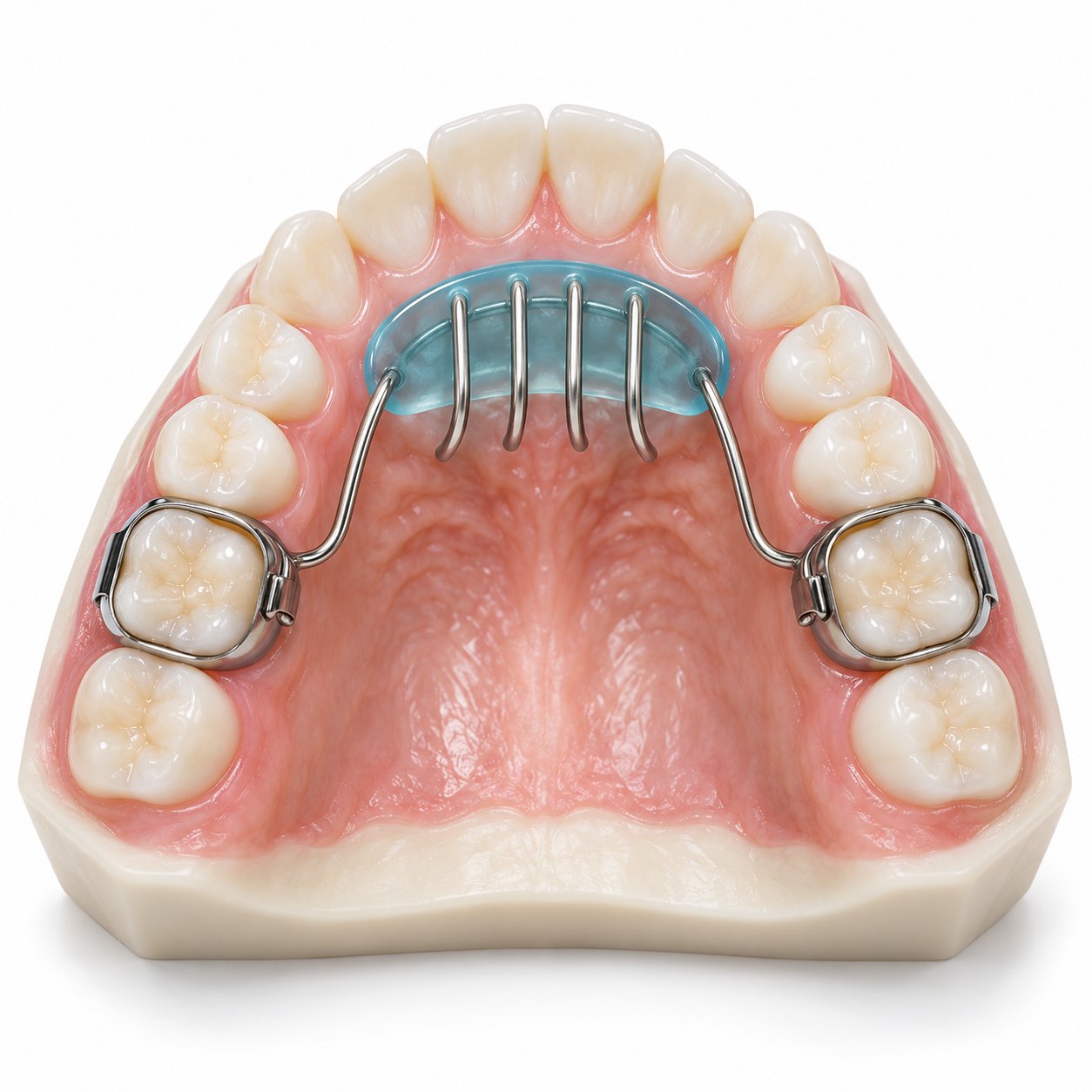
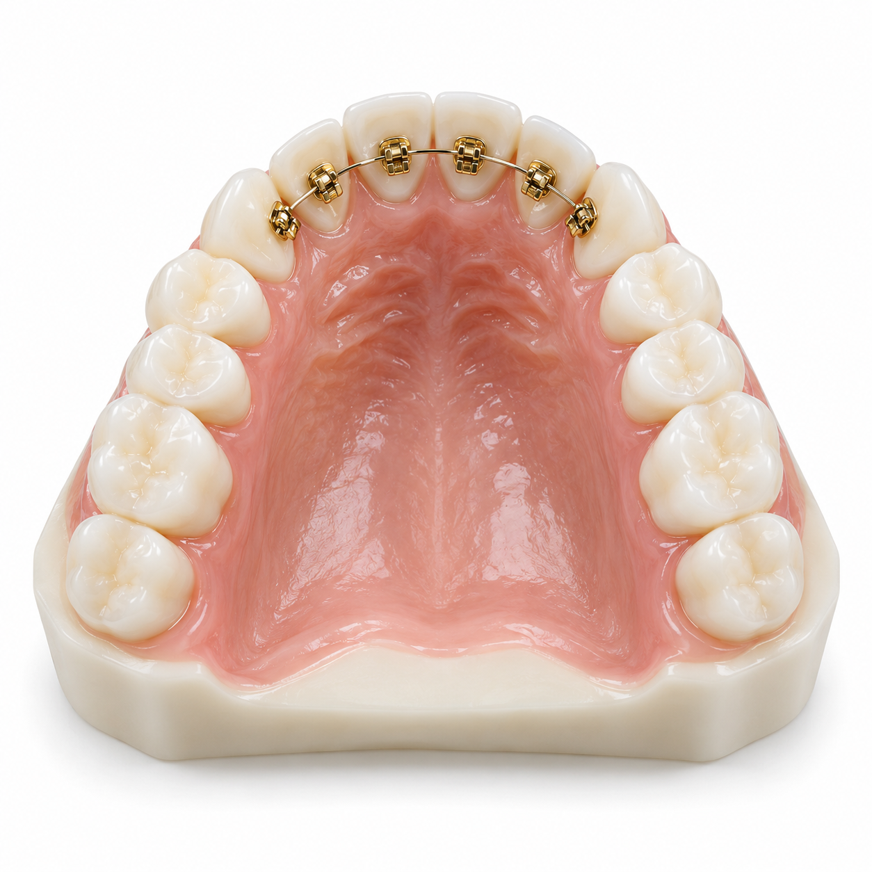
Fixed Orthodontic Appliances as Space Maintainers: A fixed appliance such as a band and loop or a lingual arch is used to maintain the open space created by missing or late-developing teeth.
Costs in Kerala vary based upon the type of treatment needed and location:
Consultation & Panoramic X-Ray: Urban clinics in Kochi, Thiruvananthapuram, and Kozhikode charge ₹800 to ₹2,500 for consultation and panoramic radiographs.
Gingival Window / Operculectomy: Minor surgical procedures are priced from ₹2,500 to ₹6,000, utilizing common diode lasers or electrosurgical methods.
Odontoma / Supernumerary Tooth Removal: Prices for odontomas/supernumeraries depend on how deep they are located within bone and/or how close they are to vital structures; prices range from ₹6,000 to ₹18,000.
Setup of Orthodontic Traction and Space Maintenance: Costs can be anywhere from ₹10,000 to ₹35,000+.
Pedodontist (Pediatric Dentist): Diagnoses and manages primary erupted malposition issues as well as the child’s soft tissues to allow for proper space management prior to eruption of the permanent teeth.
Oral and Maxillofacial Surgeon: Required for impacted third molar cases with deep bone, multiple supernumeraries that require extraction, or surgical exposure under general anesthesia (GA) or intravenous (IV) sedation.
Orthodontist: A necessity if the eruption is so severely delayed it results in a large loss of space, an arch collapse, or if continuous traction on the tooth is required.
Emergency / Urgent (Within 1 to 2 Days / 24–48 Hours): Delayed eruption causing acute pain at the site of the delay, purulent drainage, or localized swelling around the area of the delay, which would indicate the development of a pericoronal abscess.
High Priority (Within 2 Weeks): The delay is asymmetrically greater than 6 months and/or there is a retained primary tooth that has not been removed because it was still firm and will not budge even though its replacement has erupted normally.
Routine / Elective (Every 6 Months): Routine tracking of all pediatric dental patients to evaluate their growth and development during each scheduled appointment.
My child has just turned one year old, but no teeth have erupted; what is wrong with my child?
The time frame for the appearance of primary teeth varies. The first tooth usually erupts at approximately six (6) months of age. A delay until twelve (12) to fifteen (15) months is considered within normal limits. If no primary teeth are present after fifteen (15) months, a consultation with a pediatric dentist is advisable for an examination to determine the status of root development as well as to evaluate for possible systemic causes.
Is it okay to feed my baby local food such as payasam or soft rice? Will this delay the eruption of the baby's first tooth?
A diet composed primarily of very soft processed foods such as overcooked rice porridge and sweetened desserts such as *payasam* will deprive your child of the firmness required for proper stimulation of the growth of the mandible (jaw). Eating fresh, unprocessed raw fruits, vegetables, etc., along with eating firmer types of traditional foods, will provide the necessary circulation to help your child's first tooth break through from under the gum.
3. If my child's permanent tooth is late in erupting, does this mean he/she will need braces?
No. In many cases, if adequate space is maintained during childhood with the use of space maintainer appliances or small amounts of tissue removal, the permanent tooth will erupt normally. However, if the adjacent teeth move into the space left open by the missing tooth, orthodontic therapy may be indicated to allow sufficient room for the permanent tooth to come into place.
Radiographic evaluation at an early age is essential to identify any impediments to tooth development. If there are any physical barriers (supernumeraries, ectopic tooth position), these should be addressed prior to any potential impairment of jaw and facial alignment (arch deformity) and/or potential tooth impaction. The earlier the problem is identified and treated by a pediatric dentist, the less likely it will be necessary to perform invasive and expensive orthodontic treatments when your child is older.



.jpg)
.jpg)
.jpg)
.jpg)

.jpg)
.jpg)
.jpg)
.jpg)
.jpg)
.jpg)
.jpg)
.jpg)
This site is part of the DEC Dental Network — a Dental Equity & Connectivity ecosystem of clinical, educational, and patient-support brands operated by Team Dental Pvt. Ltd. Our network works as one — guiding you from your first search to your final smile.
© 2026 Team Dental Pvt. Ltd. · Founded 2013, Angamaly, Kochi, Kerala
About DEC Dental