Discolouration of the tooth occurs when there is an alteration to the translucency, colour, or hue of the dental hard structures. It is further categorized as either extrinsic staining (surface deposition onto the enamel matrix) or intrinsic staining (pulp chamber or dentinal tubule penetration with deeper discoloration). The translucent, protective layer at the outermost part of each tooth is referred to as enamel. When chromogens (pigment-bearing molecules) settle upon the surface of the enamel or enter through enamel microfractures into underlying dentinal tubules, they disrupt light transmission and refraction through the dental hard structures, thus causing a shift in the appearance of the affected tooth or teeth to yellow, brown, grey, or black.
Clinical Definition: Changes in both the translucency of the enamel and the hue of the dentin due to extrinsic staining, absorption of pigments into the dentin, or necrosis of the pulp.
Signs / Indicators That Require Urgent Care: Formation of dark brown or black structural defects associated with severe localized pain, prolonged temperature sensitivity, and/or visible gum inflammation/swelling indicating active decay or pulpal infection.
Treatment Options for Restoring Discolored Teeth: Professional ultrasonic cleaning, microabrasion, chemical vital bleaching (in-office hydrogen peroxide activation), composite bonding, and ceramic veneers.
Care Package Costs (Range): Comprehensive whitening and aesthetic restorations range from ₹3,500 to ₹22,000+ per arch, depending on the type of materials used.
Progressive Colouration: Yellow-brown to darker shades, sometimes turning blue-grey as the teeth darken over time and across the entire upper or lower dental arch.
Decrease in Translucency: As the front surfaces of the central incisors begin to lose their transparency and sparkle, they may appear duller than normal and/or have a chalky or muddy appearance.
Texture Alterations: The front of the tooth can feel rough, pitted, or like small grains to your tongue due to excessive surface plaque accumulation or enamel loss.
Darkened Individual Tooth: One specific tooth gradually turns from its original white colour to grey or completely black in the weeks or months following trauma, indicating vascular and pulpal changes occurring inside the tooth.
Stained Gingival Margin: Dark staining at the gum line bordering one side of the tooth, typically indicative of a high level of calculus accumulation, betel nut use, or tobacco tar exposure.
An immediate clinical examination is warranted if you experience sudden tooth discoloration accompanied by deep pulpal or structural distress:
Continuous Spontaneous Toothache: Persistent, pulsating pain throughout the day originating from a dark tooth, which could indicate extensive pulp tissue damage or necrosis within the tooth.
Localized Swelling: Small localized bulges or fistulae on the gums surrounding a dark tooth area, serving as evidence of a long-standing chronic abscess at the apical root end.
Delayed Post-Traumatic Color Shift: Darkening of a tooth in the weeks or months following a traumatic event or injury, indicating progressive pulpal necrosis.
Prolonged Temperature Sensitivity: Severe, sharp pain lasting for several minutes when consuming extremely hot tea, cold water, or iced drinks.
Spontaneous Bleeding: Frequent spontaneous bleeding of the gums while lightly brushing teeth in dark tooth areas, which could indicate severe periodontal disease.
Chromogen Dietary Sources: Black tea with a high chromogenic level, curries made using turmeric, drinks prepared with large amounts of berries, and coffee.
Stains from Betel Nut: Chewing paan, using tobacco, and/or smoking, which produce very heavy, difficult-to-remove dark brown and black stain deposits on teeth.
Necrotic Pulps: Traumatic injury interrupts the nerve and blood supply to the pulp tissue, causing red blood cell breakdown products to release into porous dentinal tubules.
Excessive Fluoride Use: High levels of fluoride ingested during the pre-eruptive stage of tooth development in early childhood result in hypomineralized, discolored, and pitted enamel.
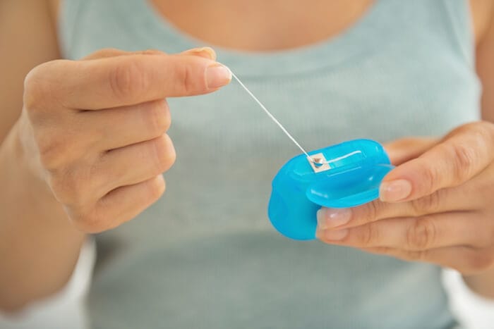
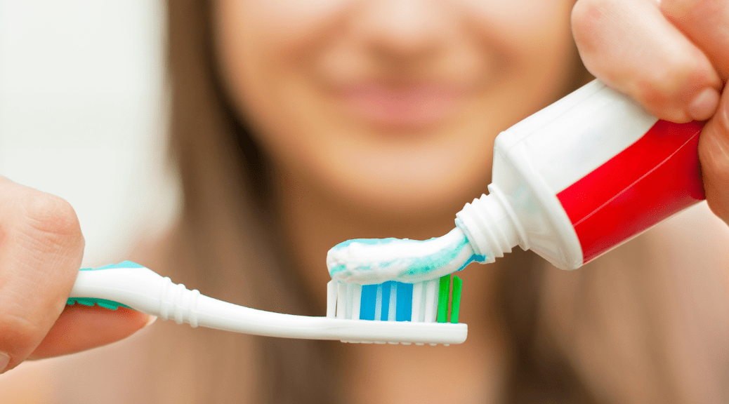
Poor Dental Care Habits: Failing to brush and floss regularly leads to the accumulation of bacteria-laden plaque and tartar, which absorbs daily food colourants.
Aging: Natural wear and tear over time thins the outer enamel layer, exposing the underlying naturally yellow-coloured dentin layer below.
Systemic Medication Exposures: Systemic ingestion of tetracycline antibiotics during pre-eruptive tooth development, causing deep intrinsic dentinal band discoloration.
Dietary Habits: Consuming large amounts of dark regional gravies, red amaranth, or spices without rinsing the mouth post-meal.
Genetic / Developmental Enamel Hypoplasia: Conditions affecting development can cause thinner, less dense enamel that is more prone to staining.
Occupation-Related Exposures: Industrial exposures to acids or drinking water with higher mineral concentrations in certain geographic regions.
A preliminary, non-invasive inspection of your mouth can be done at home using a bathroom mirror with good lighting:
Step 1 (Rinse): Swish your mouth with clean water to wash away debris lodged between and on top of your teeth.
Step 2 (Visual Inspection): Look into the mirror with bright lights shining directly onto the front and backs of each tooth. Look for small brown or black spots, overall discolouration, or an unusual colour variation on a single tooth.
Step 3 (Tactile Examination): Run your tongue along the side of the dark area. A smooth surface sensation usually signifies deeper intrinsic discoloration within the enamel or dentin, while a rough sensation usually indicates surface plaque or tartar buildup.
Step 4 (Gingiva Evaluation): Examine the gums surrounding the stained area. Are they red, puffy, or bleeding?
Step 5 (Pain Test): Tap on the affected tooth lightly with your fingertip. Do not tap forcefully. If light tapping causes significant pain, it indicates underlying periapical inflammation requiring an immediate dental appointment.
Shade Analysis: Comparing the tooth to standard dental shade charts under a controlled light source to determine the best matching shade of white or color.
Translucency Testing: Using a thin beam of light (such as fiber-optic transillumination) to pass through tooth structure and identify microcracks, enamel defects, or localized carious opacity changes.
Radiographs (Dental X-Rays): Periapical radiographs visualize internal root structures, evaluate bone density around root apices, and check for hidden cavities.
Pulp Sensitivity Testing: Using cold or electrical testing methods to verify whether the nerve inside a darkened tooth is alive, inflamed, or dead.
Staining Plaque-Disclosing Solution: Applying harmless vegetable-based dyes to reveal plaque matrices and extrinsic stain locations.
|
Grade Level |
Structural Depth |
Clinical Appearance |
Standard Clinical Pathway |
|
Mild (Extrinsic) |
Enamel Surface |
Surface yellowing, light tea/coffee film |
Professional Scaling & Polishing |
|
Moderate (Intrinsic) |
Superficial Dentin |
Generalized dark yellow, mild fluorosis specks |
In-Office Chemical Bleaching / Microabrasion |
|
Severe (Deep Intrinsic) |
Deep Dentin / Pulp |
Deep grey/brown bands, pulpal necrosis, trauma |
Endodontic Therapy followed by Internal Bleaching or Ceramic Crown Restorations |
Ultrasonic Scaling & Air Polishing: High-frequency sonic vibrations combined with fine powder streams remove superficial tartar deposits and surface stains caused by smoking or dietary habits.
In-Office Vital Bleaching: Professional high-concentration hydrogen peroxide gels activated by specialized LED lights break down and oxidize chromophores deep within the enamel and dentin.
Enamel Microabrasion: Removes small amounts of surface enamel using a mild acid and abrasives to polish out shallow white spots caused by excessive fluoride exposure.
Composite Resin Bonding: Applies composite resin directly onto a tooth to repair, shape, and re-colour a front tooth in a single office visit.
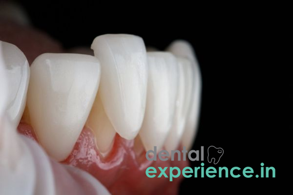
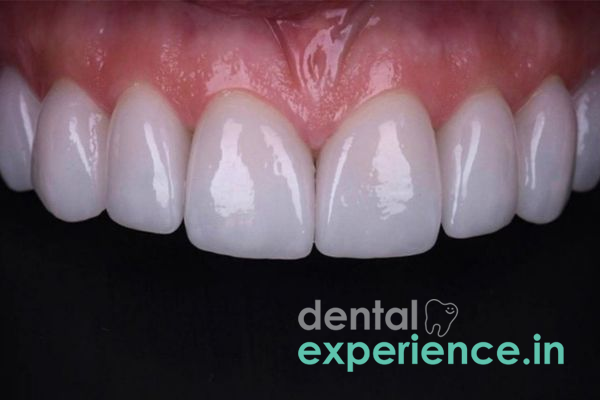
Porcelain Veneers & Ceramic Crowns: Custom-made porcelain shells bonded to the front surfaces of teeth to cover internal discolourations and restore natural translucence.
Cosmetic dental treatment costs in Kerala vary based on equipment, clinician expertise, and material selection:
Professional Scaling & Air Polishing: ₹800 – ₹2,500 per treatment
In-Office Teeth Whitening (Both Arches): ₹7,500 – ₹18,000
Direct Composite Resin Bonding: ₹1,500 – ₹4,000 per tooth
Custom Porcelain Veneer / Ceramic Crown: ₹6,000 – ₹16,000 per unit
Financial Note: Standard cosmetic whitening treatments are typically considered non-medical expenses and lack insurance coverage. However, if a root canal or protective crown is required because a tooth has died and become discolored, some insurance plans provide partial coverage for medically necessary procedures.
General Dentist: Provides all general dental services including routine cleanings, scaling and polishing, air polishing, and mild in-office bleaching.
Prosthodontist: Possesses high-level expertise in complex aesthetic dentistry, including structural ceramic veneers and full-mouth shade correction.
Endodontist: Essential when discoloration stems from nerve death or internal damage, performing precise root canals and internal "walking" bleach techniques.
Periodontist: Crucial when gum-area discoloration is linked to active infection or subgingival calculus accumulation.
Emergency (Within 24 – 48 Hours): Any sudden onset colour changes accompanied by severe pain and/or swelling of the face or jaw.
High Priority (Within 7 – 14 Days): A rapid shade change on a single tooth with no acute pain; dark bleeding around the gum line; or a rough, flaking enamel surface.
Low Priority (Elective): Mellow, widespread yellowing from food and drink consumption, cosmetic brightening requests, or general long-term stain management.
How do I explain why my front tooth turned a dark grey color weeks or months after a slight injury?
You likely experienced damage to the nerves and blood vessels in the root of your tooth during impact. Red blood cells break down inside the tooth and leak into the dentinal tubules, causing dark coloration. Visit a dentist promptly for a nerve vitality evaluation and to determine if endodontic treatment is required.
Is it effective to brush my teeth daily with herbal or activated charcoal powders to remove internal stains?
No. Homemade powders containing harsh abrasives (like activated charcoal) scratch off the outermost protective enamel layer, exposing underlying yellow dentin and making teeth even more susceptible to future staining. Intrinsic stains live deep within the dentin layer and require professional chemical oxidation or restorative bonding.
What type of risk does professional teeth bleaching pose to my natural enamel?
Teeth bleaching is safe when performed or monitored by a trained dental provider using ADA-evaluated products. Modern professional formulations include desensitizers and maintain a safe pH level that oxidizes stains without dissolving or deteriorating the enamel.
Tooth discolouration is often regarded as purely cosmetic, but clinical experience reveals it is frequently the first sign of deeper structural issues. While superficial stains from local dietary habits respond well to standard prophylactic care, localized colour changes or darker grey shades typically indicate internal pulpal compromise or developmental hypomineralization. Modern cosmetic and restorative modalities—such as high-translucency lithium disilicate veneers that preserve natural tooth structure, and vital bleaching options utilizing targeted LED technology—allow clinicians to treat patients safely and predictably. Patients are strongly cautioned against unproven home remedies, abrasive powders, and unregulated over-the-counter kits. A comprehensive evaluation process provides your dentist with the insights needed to develop a customized, safe, and reliable treatment plan.



.jpg)
.jpg)
.jpg)
.jpg)
.jpg)
.jpg)
.jpg)
.jpg)
.jpg)
.jpg)
.jpg)
.jpg)
This site is part of the DEC Dental Network — a Dental Equity & Connectivity ecosystem of clinical, educational, and patient-support brands operated by Team Dental Pvt. Ltd. Our network works as one — guiding you from your first search to your final smile.
© 2026 Team Dental Pvt. Ltd. · Founded 2013, Angamaly, Kochi, Kerala
About DEC Dental