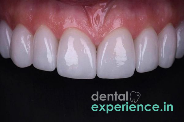
The dental condition known as "dental fluorosis" results in developmental disturbances of enamel due to excessive consumption of fluoride prior to completion of amelogenesis, taking place between the ages of 1 and 8 years for permanent teeth. During these years, developing ameloblasts (cells that create enamel) are exposed to increased levels of systemic fluoride and form an enamel matrix that has less mineral content and more protein compared to normal enamel. This can result in a range of visual effects including small white lines on teeth, large white streaks down teeth, and even extensive brown spots/pits within teeth. From a restorative standpoint, fluorosis is considered an intrinsic color alteration within the structure of the enamel itself as opposed to being merely an extrinsic stain.
Clinical Definition: Hypomineralized enamel resulting from high fluoride exposure during the early stages of tooth development before teeth erupt into the oral cavity.
Indications for Immediate Care: Brown intrinsic staining in addition to surface chipping, pits in the enamel, or hypersensitivity to cold/heat.
Common Recovery Options: Microabrasion; composite bonded restoration; ceramic veneer(s); crown coverage.
Changes in Tooth Color: White flecks on tooth, or lace-like white lines running from the gum line up into the crown area of the tooth, or wide mottled spots all over the tooth.
Staining: Intrinsic stain is dark brown to amber color which can be seen when the patient opens their mouth and will not brush off after eating a meal.
Texture of Teeth: Pits, cracks, scratches, or rough texture where there was once a smooth outer layer of enamel covering the dentin underneath.
Increased Risk of Damage: Greater risk for small micro-fractures, chips at edges, and increased sensitivity to cold foods/drinks such as cold sambharam (spiced buttermilk) or cold iced tea.
Loss of Enamel During Normal Chewing of Foods: Flakes/peel-off of enamel while eating normal foods like banana chips or roasted nuts.
Exposure of Dentin: Persistent sharp pain with consumption of either cold/hot drinks due to reduction of protective layer thickness of enamel.
Localized Caries Lesions: Food particles accumulating deep within pits created in the enamel, creating conditions for secondary decay.
Decreased Functionality: Collapse of occlusal surfaces (upper chewing surfaces) reducing function in normal chewing activity.
High-Fluoride Drinking Water: Consuming untreated borewell or deep-groundwater sources exceeding the WHO limit of 1.5 mg/L during early childhood.
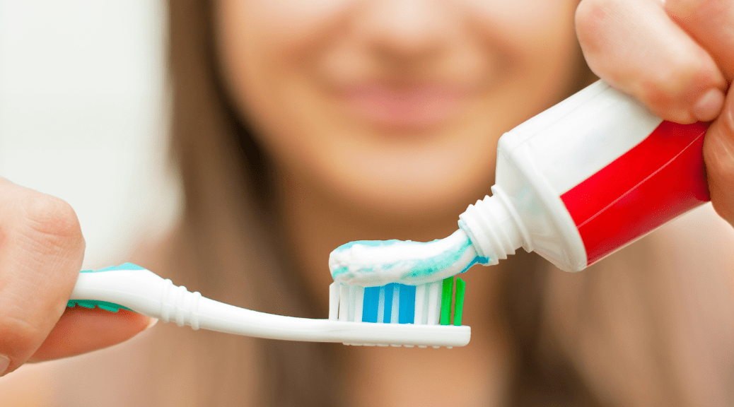
Swallowing Fluoridated Toothpaste: Ingestion of adult fluoridated dentifrices by young children under age six before swallow reflexes fully develop.
Cumulative Fluoride Sources: Combining fluoridated water supplies with dietary fluoride supplements without prior blood or water testing.
Early Childhood Exposure: Young children (under 6) who ingest an excessive amount of toothpaste containing high amounts of fluoride before they develop sufficient swallowing reflexes.
Multiple Exposures From Different Sources: Exposure to excessive amounts of fluoride through a combination of drinking water containing fluoride at levels higher than the recommended maximum concentration set by the World Health Organization (WHO) and/or ingesting dental products containing fluoride as part of a supplemental regimen for developing children.
Location-Specific Water Source(s): Relying on unregulated, deep-well water in areas where naturally occurring high levels of fluoride have been found in the groundwater of Kerala, India (such as Palakkad and Alappuzha).
Child Developmental Stage: Permanent tooth development taking place in the jaw below the surface of a child's mouth between the ages of one and eight.
Food Type Consumption Patterns: A diet consisting of food prepared using water with very high levels of fluoride or consuming large quantities of teas made with natural fluoride-rich tea leaves.
Perform a preliminary self-check prior to a clinical visit:
Clean & Dry Your Teeth: Clean your hands completely, then take a seat in a well-lit area with a clean bathroom mirror. Carefully dry the front side of the teeth with a piece of paper towel; fluorosis stains will be visible once they are dry.
Inspect the Color: Look for areas that have a flat white opaque line or brown spots that are hard to remove.
Tactile Check: Place the tip of your clean tongue or finger gently along the edge of each tooth to see if there is an uneven surface or any sharp points.
Important Safety Warning: Do not use metal instruments or sharp objects to scratch or probe your teeth.
Visual Inspection & Transillumination: A dentist uses very bright light passed through optical fiber strands to distinguish between hypomineralization of the enamel layer of a tooth and other forms of decay such as early white spot caries.
Dean's Fluorosis Index: A standardized clinical method of determining the extent of the damage caused by fluoride by scoring the two most severely affected teeth.
Radiology (X-Rays): Digital bitewing and periapical radiographs to determine the amount of dentin present, the depth of the pulp chamber, and to eliminate proximally located decay.
Vitality Testing: Thermal or electrical pulp testing on severely pitted teeth to verify endodontic vitality.
|
Grade Level |
Clinical Presentation |
Enamel Condition |
|
Very Mild |
Slight white aberrations or small white flecks covering a minimal area of the surface |
Intact mineral structure |
|
Mild to Moderate |
Opaque white bands or brown staining covering up to 50% of surface |
Smooth to slightly rough surface |
|
Severe |
Marked brown discoloration, extensive surface pitting, corroded appearance |
Structural loss, prone to chipping |
Enamel Microabrasion: Using 6.6% to 10% hydrochloric acid slurries (containing silicon carbide micro-particles) that gently remove enamel containing white or brown staining (up to 100 microns).
Resin Infiltration: Low-viscosity resins enter into hypomineralized enamel pore spaces after etching, providing a color match to healthy enamel without requiring tooth preparation like traditional fillings.
Direct Composite Veneering: The direct bonding of nano-hybrid composite resins onto stained enamel surfaces provides immediate masking of moderate staining and restoration of contour defects within a single appointment.
Ceramic E-Max / Zirconia Veneers: Custom laboratory fabrication of ceramic facings. Porcelain provides excellent aesthetic results and outstanding durability when used on severely discolored teeth.
Full Coverage Crowns: Due to extensive enamel loss from pits and/or erosion, monolithic zirconia or porcelain-fused-to-metal crowns are reserved for patients requiring full coverage restorations due to severe structural damage.
|
Procedure |
Cost Range (INR / ₹) |
|
Microabrasion / Resin Infiltration |
₹2,500 – ₹5,000 per tooth |
|
Direct Composite Bonding |
₹2,000 – ₹4,500 per tooth |
|
Ceramic Veneers (E-Max / Zirconia) |
₹10,000 – ₹22,000 per tooth |
|
Full Ceramic Crown |
₹8,000 – ₹18,000 per tooth |
General Practitioner: Handles initial diagnosis, Dean's index grading, preventive counseling, and simple microabrasion or direct composite bonding.
Prosthodontist: Essential for complex full-mouth rehabilitation, severe brown stain coverage using ceramic veneers, and smile design.
Pediatric Dentist (Pedodontist): Manages early-stage fluorosis, enforces preventive water-swapping strategies, and treats young children.
Conservative Dentist & Endodontist: Specializes in resin infiltration, indirect esthetic restorations, and pulp protection for deeply pitted teeth.
Elective (Scheduled within 2–4 weeks): Mild white flecks or cosmetic brown discoloration without physical enamel breakdown.
Prompt (Scheduled within 3–7 days): Rough surface pitting causing persistent food impaction or mild thermal sensitivity.
Urgent (Within 24–48 hours): Fractured enamel edges, severe sensitivity to hot/cold liquids, or localized soft tissue inflammation caused by sharp tooth borders
How do you tell whether a child has fluorosis or an early case of dental caries (tooth decay)?
If your child has fluorosis, affected teeth will be involved symmetrically across both sides of the mouth. Fluorosis produces uniform intrinsic white bands or brown spots that appear as the teeth erupt. An early case of dental caries (demineralization) occurs first at the gumline or in chewing grooves as small, soft, sticky white spots where plaque bacteria accumulate and produce acid.
Is drinking boiled water enough to keep fluoride away from my children?
No. Boiling water does not remove fluoride; as water evaporates during boiling, the fluoride concentration actually increases. If your family uses high-fluoride well water (common in parts of Palakkad and Alappuzha districts in Kerala), consider installing a Reverse Osmosis (RO) system or using activated alumina fluoride-removing filters.
Will tooth-whitening products remove all dark-brown spots of fluorosis on teeth?
Tooth whiteners may lighten surrounding enamel to make fluorosis less noticeable, but they almost never eliminate deep, dark-brown fluorosis spots. Professional microabrasion, resin infiltration, composite bonding, or porcelain veneers are required to cover or eliminate deep intrinsic staining.
Fluorosis continues to be a major focus of dental public health efforts within areas of Kerala that have high levels of geologically derived fluoride in their groundwater sources exceeding optimal concentrations. Dental clinicians must adopt a minimally invasive approach when managing this condition. The best time for early diagnosis of dental fluorosis is during pediatric development stages, allowing for easy removal of superficial stains via microabrasion and/or resin infiltration of affected areas to preserve tooth structure. Adults who present with moderate to advanced brown stain and surface porosity secondary to fluorosis can achieve long-term aesthetic restorative success using modern indirect ceramic systems (such as lithium disilicate) while providing protection against dentin exposure. Community prevention actions — specifically transitioning to RO-filtered drinking water prior to early childhood amelogenesis — remain the most effective method for eliminating the risk of developing fluorosis.



.jpg)
.jpg)
.jpg)
.jpg)

.jpg)
.jpg)
.jpg)
.jpg)
.jpg)
.jpg)
.jpg)
.jpg)
This site is part of the DEC Dental Network — a Dental Equity & Connectivity ecosystem of clinical, educational, and patient-support brands operated by Team Dental Pvt. Ltd. Our network works as one — guiding you from your first search to your final smile.
© 2026 Team Dental Pvt. Ltd. · Founded 2013, Angamaly, Kochi, Kerala
About DEC Dental