Food lodged in gaps—clinically referred to as an "interdental food impaction"—occurs when a particle of a firm food item is stuck in the interproximal space between two adjacent teeth. When teeth are healthy, tight interproximal contact points prevent food from wedging between them and protect the interdental papilla.
Disruptions can occur when there is decay on one or both of the adjacent teeth, causing a disruption of their normal contact; when there has been significant loss of supporting bone around the roots of adjacent teeth resulting from periodontal disease; when restorative materials do not fit properly on one or both of the adjacent teeth; or when gums naturally recede with age and lose their ability to cover and protect the roots of teeth.
When left untreated, impaction mechanically causes a local environment that allows bacterial growth which produces acids leading to demineralization and carious breakdown of enamel, continuous chronic inflammation of the gums, and rapid loss of supporting bone of the alveolus.
Clinical Definition: Interproximal contact impaction is when food becomes impacted between two teeth because of the lack of sufficient contact between the surfaces of the adjacent teeth, or as a result of recession of the gingiva, or as a result of movement of the teeth apart.
Emergency Signs: Sudden throbbing pain, pus oozing through the gums, constant bad breath, bleeding after light pressure, or a sudden separation of adjacent teeth.
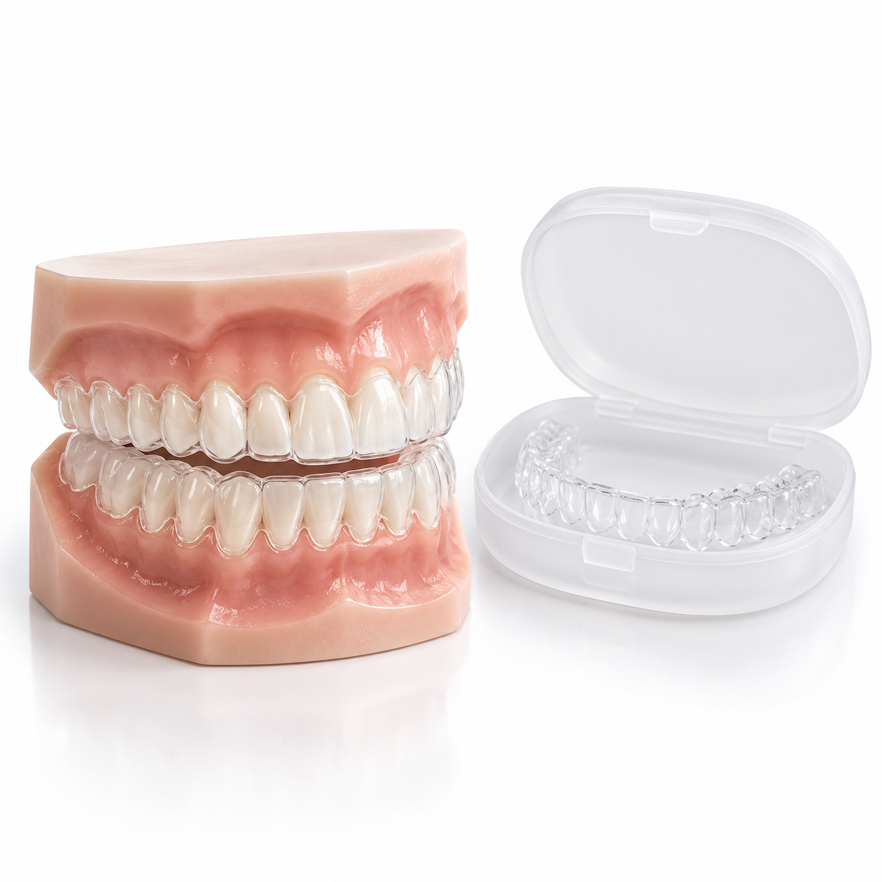
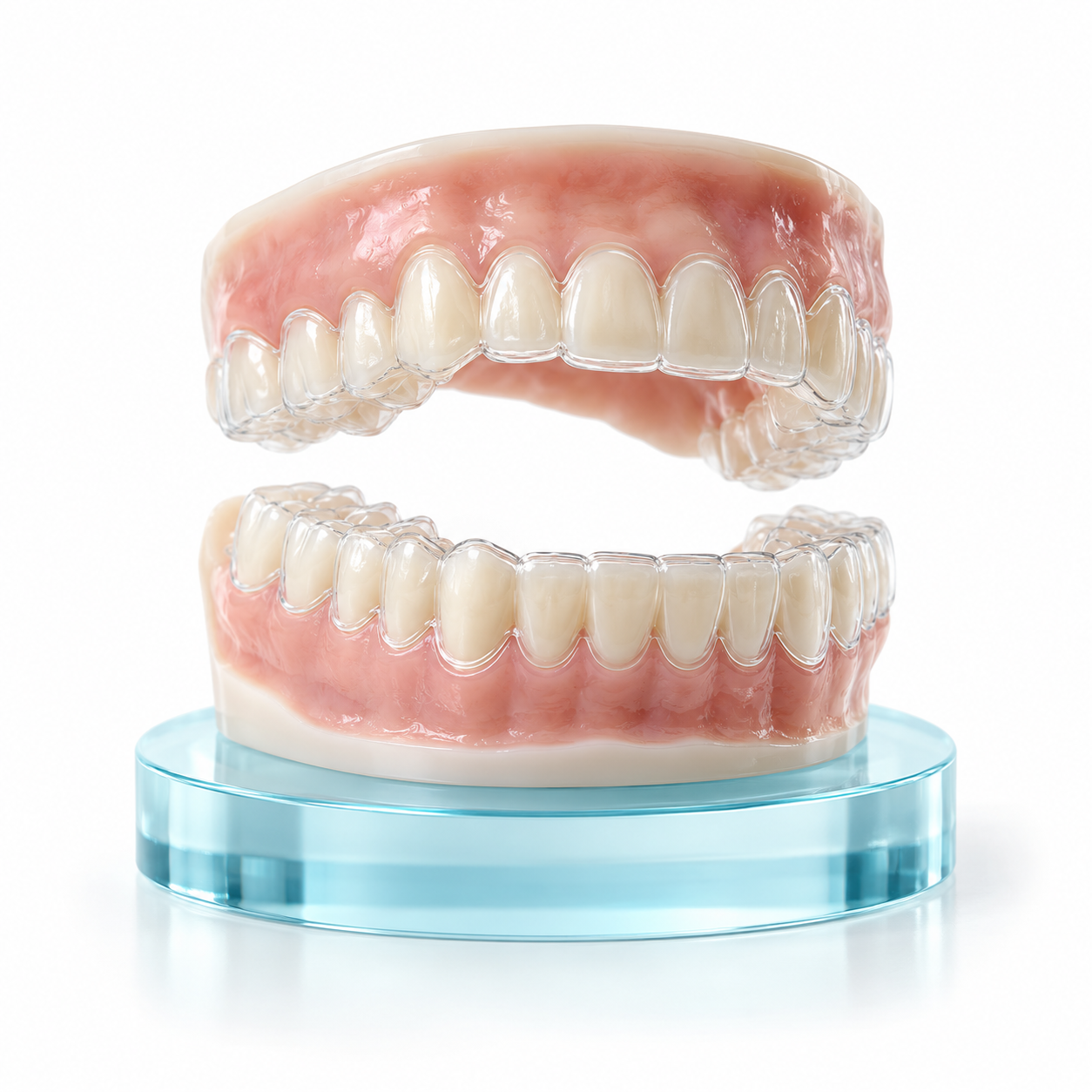
Treatments: Bonding composite resins to restore the missing contact area, manage periodontal diseases affecting the interdental tissues, attach custom porcelain veneers to create new contact areas between adjacent teeth, create CAD/CAM crowns for severely damaged or weakened adjacent teeth, or use orthodontic treatment using transparent removable appliances to move adjacent teeth back closer together.
Food impaction symptoms are both physically and sensitively noticeable to patients:
Tactile Pressure: Tactile (touch) pressure between two teeth immediately following ingestion.
Interdental Soreness: Dull ache in the gums surrounding the impacted area which usually resolves once the impacted food is dislodged with force or removed using floss.
Gingivitis: Erythema (redness), edema (swelling), bleeding at the site of the impacted food item while brushing/flossing around the affected area.
Halitosis: Persistent bad breath/sour taste resulting from anaerobic bacterial decomposition of retained food/organic matter.
Thermal Sensitivity: Brief pain upon contact with hot/cold objects as soon as pressure is released and/or dentin is exposed or root surface(s).
Continuous Throbbing Pain: Indicates hyperemia of the pulp chamber and/or inflammation secondary to deep interproximal caries.
Gum Abscess Formation: Local, red, swollen, fluid-filled bumps on the gum line indicating significant deep periodontal pocket involvement.
Significant Recession of Papillae: Rapid development of large "black triangles" due to alveolar bone loss.
Tooth Mobility: Noticeable movement/drifting of adjacent teeth under normal bite forces.
Food lodgment is caused by numerous mechanical, structural, and behavioral aspects of tooth form and function:
Interproximal Decay: The presence of an unaware interproximal decay in the side surface of one or both adjacent teeth can lead to disruption of the smooth contact point between those two teeth. This creates a space for food to lodge.
Gingival Recession and Periodontitis: Chronic gum disease can cause destruction of the support structures of the tooth such as the periodontal ligaments and surrounding bone. As a result, the gums will begin to recede and expose wider spaces around the roots of the teeth.
Defective Dental Restorations: Defective restorations may be overhanging, worn out, or improperly shaped so they do not fit closely enough against each other at the interproximal area. Additionally, some defective restorations are missing their interproximal contact areas altogether.
Tooth Pathologic Migration: A missing tooth (particularly a missing molar) causes the adjacent teeth to tip and migrate mesially into the space or allows opposing teeth to super-erupt occlusally, increasing the amount of space between the teeth and making it easier for food to get trapped.
Mechanics of Diet: Tough, fibrous regional diets (such as dried meats, coconut fibers, etc.) that have a tendency to act like wedges when subjected to heavy biting forces can force food into the interproximal space between the teeth.
Untreated Periodontal Disease: The chronic inflammatory process associated with untreated periodontal disease causes weakening of the alveolar bone matrix.
Aging: Gingival recession due to aging as well as the normal wearing away of enamel over time contributes to increased food lodging.
Poor Oral Hygiene: Failure to remove plaque from the interproximal areas leads to inflammation of the interproximal tissues and subsequent loss of these tissues and thus further increases the risk of having lodged food.
Crowding/Malocclusion: Malaligned or crowded teeth result in irregularly shaped contact points between them. These irregularities increase the likelihood that a piece of food will become lodged in this region.
Use this self-assessment process to find out about your food retention zones safely. Do not harm your delicate gum tissues!
Visual Assessment: Use a mirror under bright light. Look for an area that has space, "black triangles," or debris from food between your teeth.
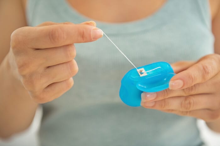
Floss Test: Carefully guide an unwaxed dental floss through the contact point you believe may have a gap.
Resistance Test: When you drop the floss down easily into the gap without hearing a gentle snapping sound, or when the floss cuts like a knife (shatters), then your interproximal contact is either loose or rough.
Tissue Test: See if the gum tissue between those two teeth bleeds at once, or if they appear darker red and are swollen compared to healthy, pink gums.
Safety Warning: Never use metal pins, sharp needles, or hard wooden sticks to get trapped food out of a contact point gap—this will cause irreversible damage to the gum and bone that connect it.
Your dentist performs an examination and makes a diagnosis based on several steps:
Visual & Tactile Probing
Examination with a fine explorer probe.
The dental professional inserts a shimstock foil (typically 8 to 12 microns thick) or dental floss and evaluates the tightness of the contact points.
Periodontal Probing
The dental professional measures the depth of the pocket created by the gums around each tooth. This measurement is made using a calibrated periodontal probe.
The measurements are taken to determine the health status of your gums.
The deeper the pocket, the greater risk for severe gum disease.
Digital Radiographs
The dental professional takes intraoral bitewing x-rays. These x-rays show hidden areas of decay between teeth, overhanging fillings, and the amount of bone lost due to severe gum disease.
Pulp Vitality Testing
The dental professional checks to make sure that the nerve inside each tooth remains vital.
|
Severity Level |
Clinical Presentation & Characteristics |
|
Mild (Superficial) |
The occasional food gets caught in this space, but there is no bone loss and the interdental papilla is still intact. There may be some temporary discomfort that can usually be resolved by using waxed floss. |
|
Moderate (Structural) |
Daily food gets stuck in the area. Gingivitis develops along the gums on one side, as well as black triangle areas where food accumulates. Deepening of the pocket for a moderate amount of time (between 3–5 mm). Early stage development of interproximal enamel decay. |
|
Severe (Complex) |
Constantly large amounts of food get impacted into the space. Large amounts of destruction occur to the supporting structures of your teeth. Pockets are extremely deep (over 5 mm) and/or you experience excessive tooth movement. Potential for developing an abscess due to infection. |
Using modern restorative options available today allows dentists to create long-lasting, precision corrections:
Direct Composite Restorative Dentistry: High-strength nano-hybrid composite resins used to attach to the surface of each tooth to fill small gaps completely, without need for follow-up appointments.
Computer-Aided Design / Computer-Aided Manufacturing (CAD/CAM) Dental Crown & Inlay/Onlay Restorations: Custom-made ceramic or zirconia restorations that have been milled with accuracy to fit snugly back into place to restore anatomically correct, tight contact points between adjacent teeth.
Deep Interdental Periodontal Therapy: Specific specialized treatments for treating gum infections in the deeper spaces around the roots of teeth which will help repair the destroyed tissue and stabilize loose teeth.
Orthodontic Treatment Using Clear Aligners: Digital clear orthodontic appliances designed to move individual teeth out of their current position and into proper alignment to eliminate gaps throughout the entire dental arch.
The cost of treatment in Kerala will depend on several factors including how complex the clinical issues are, what type of materials are used (for example, composite resin), and the equipment at hand within the dental clinics:
Gap Closure with Composite Resin: ₹1,500 to ₹3,500 per surface area of tooth.
CAD/CAM Ceramic Crown/Porcelain Veneer: ₹8,000 to ₹18,000 per unit.
Deep Scaling & Root Planing: ₹2,000 to ₹5,000 per quadrant.
Clear Aligner Treatment: ₹60,000 to ₹1,50,000 per case, depending on the severity of the case.
Most dental services provided privately are an out-of-pocket expense. The financial framework is as follows: Tertiary Medical College Hospitals or Government Dental Clinics throughout Kerala provide a lower-cost option for periodontal care, whereas some private health insurance add-on policies provide coverage for medically necessary restorative services.
General Dentist: Qualified for most of the routine dental work (simple fillings, composite bonding, etc.) but will refer you to a specialist if your case requires advanced treatment.
Prosthodontist: When you require something that can be made with porcelain (like a crown) that also has to look perfect in terms of color, shape, and size. They are also qualified to do extensive restoration procedures like all-ceramic bridges or implants.
Periodontist: If your gum disease has caused significant damage such as the presence of large amounts of loose gum tissue, lost teeth, or exposed roots; these are symptoms of periodontal disease.
Orthodontist: You need an orthodontist if your teeth are too far apart and your upper and lower jaws don't fit together properly.
Emergency Care (Within 24 hours)
Acute severe throbbing pain
Trauma-induced sudden tooth gaps
Pus drainage
Sudden swelling of gums/faces
Routine Care (Scheduling within 1–2 weeks)
Food gets stuck daily
Mild local soreness due to food getting stuck
Recurring bleeding when you floss
Noticeable gaps w/o immediate acute pain
Is it okay to use normal wooden toothpicks to remove food from between your teeth?
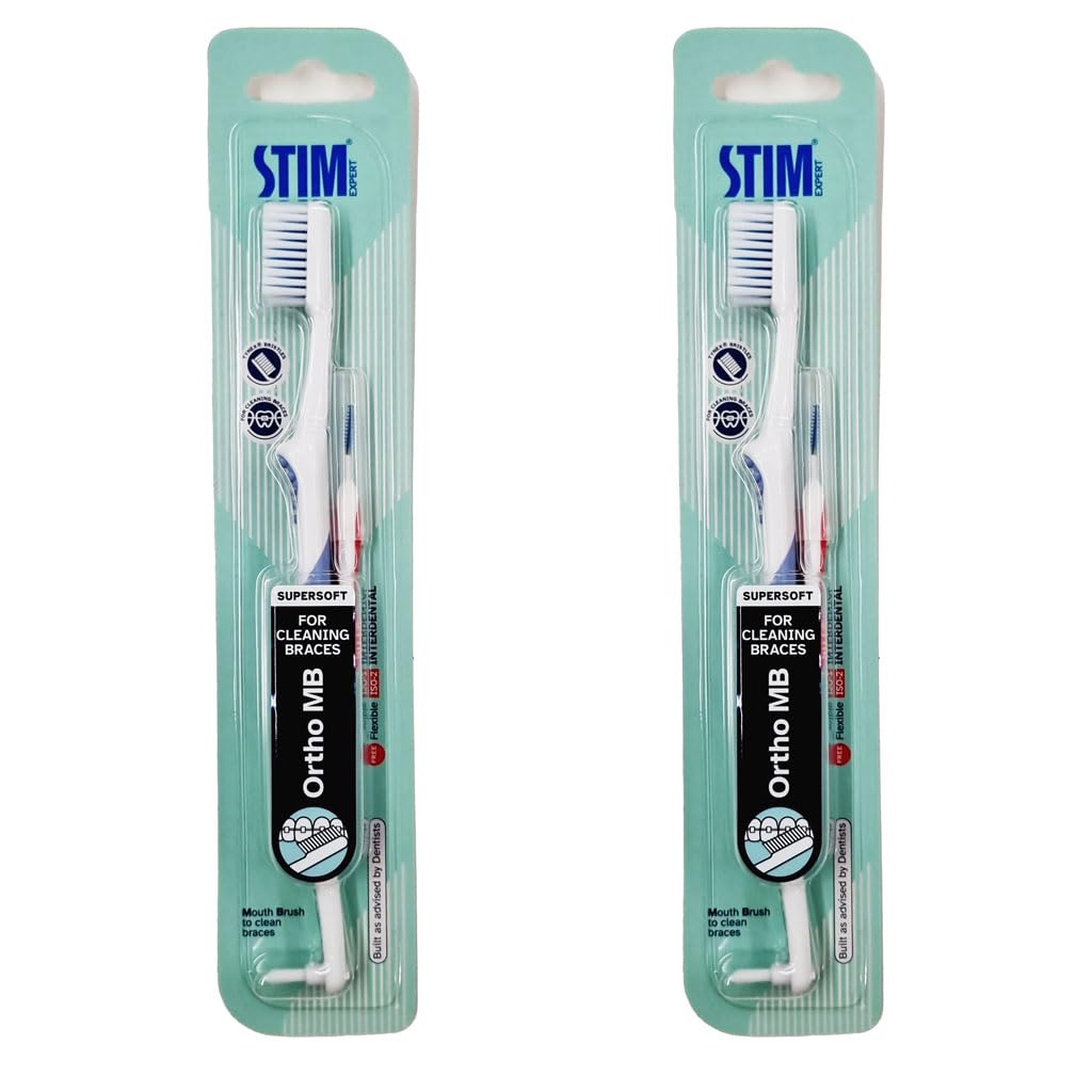
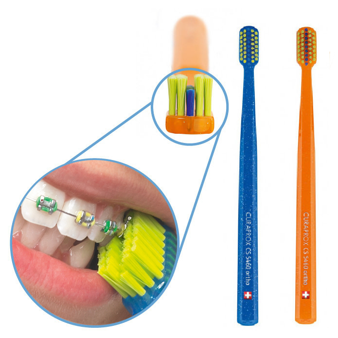
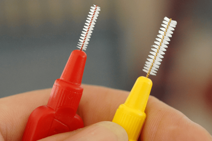
No. Wood picks are hard and will cause blunt trauma that will cause the interdental papilla to be damaged over time and make the space wider. Rubber tip interdental brushes or water flossers are recommended for cleaning between the teeth.
Won't composite bonding on top of my natural enamel hurt my enamel?
No. Direct composite bonding is a conservative, minimally invasive procedure that preserves natural tooth structure. While minimal or no enamel removal is required, the underlying surface is lightly etched with acid to ensure a strong adhesive bond.
How come after years of having no spaces in-between my teeth, now I have problems because food keeps getting caught in spaces?
Spaces often indicate gum recession, age-related tooth movement, potential cavities that occur between teeth, or an already loose existing dental restoration.
Food lodgement in interproximal spaces is more than just an annoyance to patients with existing interproximal pathology; it can serve as a clear indicator of active periodontal disease and/or continued or worsening structural compromise. The process of inserting foreign materials into compromised areas repeatedly initiates the inflammatory process (i.e., gingivitis) and significantly accelerates the rate of bone loss. Early diagnosis by a trained dental provider utilizing diagnostic technology enables treatment options that are both conservative and minimally invasive, allowing for the preservation of natural tooth anatomy while protecting the patient's long-term oral health and supporting gums.


.jpg)
.jpg)
.jpg)
.jpg)
.jpg)
.jpg)
.jpg)
.jpg)
.jpg)
.jpg)
.jpg)
.jpg)
This site is part of the DEC Dental Network — a Dental Equity & Connectivity ecosystem of clinical, educational, and patient-support brands operated by Team Dental Pvt. Ltd. Our network works as one — guiding you from your first search to your final smile.
© 2026 Team Dental Pvt. Ltd. · Founded 2013, Angamaly, Kochi, Kerala
About DEC Dental