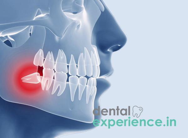
An impacted third molar develops when the third molar is blocked from emerging into the mouth either by bone, by gum, or by one or more adjacent teeth. When an impacted third molar grows at an unusual angle to the surrounding teeth and/or to the supporting jawbone, the normal anatomical arrangement of the teeth in the back of the mouth is disrupted. This disrupts the structural integrity of the posterior portion of the mouth and exerts a constant force upon the surrounding structures. This pressure triggers pathological external root resorption, where the body's cells gradually dissolve the protective cementum and underlying dentin of the adjacent second molar's root. Left unchecked, this destructive process can permanently compromise the structural integrity of the neighboring tooth.
Definition: Third molar that cannot emerge into the mouth due to lack of space.
Symptoms: Radiating jaw/ear pain, tightening (trismus) of the jaw, facial swelling, pus, and pericoronitis.
Treatment Options: Surgical extraction, operculectomy (flap removal), and orthodontic alignment with surgery
There are several physical, tactile and visual signs associated with an impacted third molar.
The most common sign is a swollen, reddened and inflamed gum flap (operculum) which covers the area where the third molar is located. Food and bacteria accumulate under this flap creating a perpetual unpleasant taste sensation and extreme bad breath.
There is also typically a deep dull ache in the jaw that worsens when you chew and radiates upward to the ear and temple. It may be possible to see a small white nub of tooth crown sticking through the gum flap.
Over time, natural jaw maturation and the normal tendency of human teeth to drift forward can cause the front (anterior) teeth to become crowded, twisted, or shifted—a natural alignment change that can be further complicated by localized wisdom tooth pressure.
Trismus: Sudden, severe restriction of jaw movement where you cannot fit two to three fingers vertically between your front teeth, indicating the infection has spread to the masticatory muscles.
Swelling beyond the jawline: Visible Swelling beyond the confines of the mandible including cheeks or neck areas indicative of facial space infection or cellulitis.
Ongoing pus drainage: Persistent pus draining from the gum margin combined with a fever greater than 100.4°F (38°C).
Severe Bite Pain: Sharp, localized pain when biting down—even on soft foods—is typically a sign of deep structural decay (root caries) forming on the hidden backside of the second molar, or severe inflammation of its surrounding periodontal ligament.
Evolutionary mismatch: Changes from consuming raw foods to soft processed foods have resulted in smaller human jaws. As a result, there is less space in the human jaw to accommodate all 32 teeth.
Misaligned mechanical forces: Developing follicle angulation anomalies cause teeth to grow forward into the adjacent second molar's roots instead of growing vertically.
Traditional dietary impact in Kerala: Sticky foods like ada, ari murukku and tapioca trap food particles under the gum flap allowing bacteria to accumulate and exacerbate pain.
Jaw size / shape: Smaller jaw size / shape Limits space behind second molars making impaction likely.
Age: Risk peaking between 17 – 26 years old during normal eruption times; after 30 years of age, bones solidify limiting access for surgeons.
Local chronic inflammation: Poor management of type 2 diabetes compromises systemic immune function lowering local oral immunity increasing frequency of episodes of pericoronitis.
Perform a safe visual self-assessment at home without irritating tissue further
Steps:
1. Sanitize your hands completely to prevent introducing additional bacteria into the mouth.
2. Utilize flashlight from smartphone to illuminate area behind chewed-on molars.
3. Gently pull the cheek outward using a clean tongue depressor or smooth back of the spoon.
4. Assess visual changes: look for asymmetric red flap of gum, partial visibility of white crown poking out at angle, slight Fluid ooze.
5. Assess tactile pressure: apply gentle pressure on the outside of the lower jaw bone near the angle of mandible. Identify deep bruise aches and/or warmth compared to the opposite side. Avoid probing areas with Sharp objects like tooth picks.
Objective evaluation by dentist requires advanced imaging tools
Imaging techniques used include:
Panoramic radiographs (OPG): Wide-angle X-ray to show exact angle and depth of tooth relative to main nerve of lower jaw.
Cone beam CT scans (3D): Maps exact space between tooth roots and vital nerves where standard X-rays show them touching.
Probing/vitality tests: Determines the amount of fluid pocket around the second molar and uses a temperature test to determine the health of the inner nerve.
|
Graded Level |
Clinical Presentation |
Structural Impact |
|
Position A: Superficial |
The chewing surface of the wisdom tooth is at or above the level of the neighboring second molar's crown. Covered primarily by a soft tissue flap. |
High risk for localized pericoronitis (gum infection); traps food debris easily. |
|
Position B: Moderate |
The chewing surface is partially trapped below the gum line and sits halfway down the crown of the adjacent second molar. |
High risk of accelerating root decay or bone loss on the backside of the second molar. |
|
Position C: Deep |
The wisdom tooth is completely embedded deep within the jawbone, sitting below the gum line and often near the main lower jaw nerve. |
Can trigger the formation of dentigerous cysts; high risk of dissolving the neighboring tooth's roots. |
Resolving an impacted third molar requires targeted minor oral surgery based upon degree of impaction
Types of surgical procedures include:
Minor surgical disimpaction: Removes blocking bone layer using precision handpiece through small gum incision. Section teeth into smaller pieces for gentle removal protecting nearby nerves and second molars before site is stitched together using dissolvable thread.
Operculectomy: A laser or electrosurgical tool is used to trim away the inflamed gum tissue flap covering a partially erupted tooth. This is highly effective only if the jaw has adequate physical space and the tooth has a normal, straight path of growth. If the tooth is structurally or osseously (bony) impacted, the gum tissue will likely grow back, making extraction necessary.
Dental treatment costs vary across Kerala depending on complexity of impaction and clinic location
Cost estimates for services offered in Kerala are:
Diagnostic work up (OPG/xray or CBCT scan): ₹800 to ₹3500.
Routine Extraction (Simple): ₹2500 to ₹4500 per tooth.
Complex Surgical Disimpaction (including bone cutting): ₹6000 to ₹15000 per tooth for surgery in major cities like Ernakulam/Kozhikkode.
General Dentists: Ideal for simple straight line extractions where teeth can be seen clearly and do not require any bone cutting.
Oral and Maxillofacial Surgeons: Necessary for more complicated cases which involve deep bone impacts especially those involving wrapped lower jaw nerves due to longer roots; also provides safety against damage to surrounding nerve structures during procedure.
Emergency window (2-4 hrs): Visit emergency room immediately if breath difficulty exists; neck/eye swelling; fever above 100.4° f (38° c); trismus due to possible life-threatening infection such as Ludwig's Angina.
Urgent Care Window (24-48 hrs): Visit dentist within two days for painful chewing; bad taste; pus emission; halitosis to prevent spread of acute infection.
Elective window (2-4 wks): Schedule a preemptive extraction if the tooth is currently pain-free but clinical X-rays show it is growing forward (mesially), trapped against the second molar, or preventing proper hygiene at the back of the mouth.
How long does average recovery take following wisdom tooth extraction?
Average time frame for healing of gum tissue after extraction is 7 - 10 days (requires soft foods and rest); time frame for rebuilding of jawbone beneath extraction site is approximately 3 - 6 months.
Will untreated impacted wisdom teeth cause my front teeth to become crooked?
Not entirely. While they can exert localized pressure, modern orthodontic consensus attributes late teenage front-tooth crowding mostly to natural jaw maturation and forward dental drift, rather than purely the "pushing" force of a third molar.
Is the surgical extraction process painful?
No. The procedure is pain-free due to use of local anesthesia/sedation. Mild soreness/swelling lasts for 3 - 5 days post-surgery and manageable with prescribed medications.
Prevention is key when dealing with third molar impaction; waiting until late 20s or early 30s increases risk since tooth roots continue to grow longer potentially wrapping around the lower jaw nerve, and surrounding bone becomes denser making it more difficult for surgeons to cut safely through the bone without damaging the nearby nerve. Removing problematic wisdom teeth between ages 18 - 22 protects second molars, preserves teeth alignment.
Postoperative care includes no smoking, avoiding straws and eating cool soft foods like yogurt rice for the first few days to prevent blood clot healing.














.jpg)
.jpg)

.jpg)
.jpg)

.jpg)
.jpg)
.jpg)
.jpg)
.jpg)
.jpg)
.jpg)
.jpg)