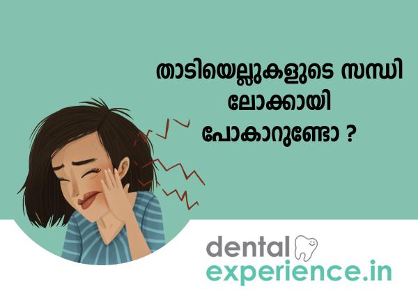
Acute temporomandibular joint (TMJ) dislocation — clinically referred to as "locked jaw" — is an emergency condition affecting the masticatory system. The TMJ connects the mandible (lower jawbone) to the temporal bone of the skull and acts as a sliding hinge allowing for rotational and translational movement. An articular disc consisting of fibrocartilage exists between these bones providing cushioning for impact and ensuring smooth movement. A locked jaw can occur in one of two forms: open-lock (anterior subluxation or dislocation where the condyle becomes lodged anterior to the articular eminence) or closed-lock (where an anteriorly displaced articular disc fails to reduce during translation, physically obstructing the condyle from moving forward and significantly restricting mouth opening). In the context of TMDs, when a patient develops a locked jaw they have transitioned from a functional balance of their TMJ into a mechanical obstruction. This needs to be addressed promptly to prevent further elongation of the ligaments surrounding the joint and subsequent damage to the TMJ capsule.
Clinical Definition: When the temporomandibular joint becomes mechanically immobilized either through displacement of the articular disc or through dislocation of the condyles.
Indications For Immediate Treatment: The ability to suddenly shut or open your mouth, unevenness in how the lower jaw closes against the upper jaw, extreme pain in front of your ears (preauricular), and sudden tightness of your masseter muscles.
Treatment Options: Clinical manual reduction of the joint, use of occlusal splints (night guards), arthrocentesis (washing out of the joint space), injection of botulinum toxin into affected muscles to allow for relaxation of those muscles, and surgical repair of the joint (chronic/ degenerative).
Locking of the TMJ can be identified with the following clinical features:
Incomplete Opening Of The Mouth: In cases where the joint has "locked" patients are unable to open the mouth to more than 20 mm. (Normally the mouth will open approximately 35 – 50 mm.)
Open-Lock: Patients who have a "locked" jaw are unable to bring the upper and lower teeth together.
The "Pop": A sudden cessation of the normal audible click associated with the articular disc as it moves back and forth when chewing or speaking usually occurs before a closed-lock and indicates that the disc has been totally dislodged from its correct anatomical position.
Pain/Tenderness Preauricularly: This pain is sharp, localized and located directly in front of the tragus of the ear. It may also radiate to the area of the temple, neck and angle of the mandible.
Asymmetrical Jaw Movement: When an attempt is made to move the jaws apart, the mandible appears to veer or sway towards the side of the locked joint because one condyle is limited in its range of motion.
Muscle Rigidity/Spasms: There is intense muscular rigidity/spasm present in both the masseter and temporalis muscles when these muscles contract to maintain the stability of a compromised joint.
Immediately evaluate your patient clinically if you note red flag indicators of possible neurologic compromise, vascular injury or tissue necrosis:
Inability to control oral secretions or swallowing saliva.
Swelling around the face/mandible, heat and/or erythema rapidly progressing.
Numbness/tingling sensation in either cheek, lower lip, or tongue (paresthesia in all three divisions of the trigeminal nerve).
Systemic signs such as fever, chills, etc., along with marked stiffness of the TMJ, suggestive of septic arthritis.
Locked jaw occurring immediately after a direct high impact trauma to the mandible or jaw, indicative of a fracture of either the mandible or the neck of the condyle.
Mechanical Displacement Of The Intra-Articular Disc: The intra-articular disc is displaced anteriorly from its normal position at rest; it acts like a doorstop to block the movement of the mandible's condylar process.
Forceful Over-Extension: Forceful yawning, protracted max. mouth opening with long-term dental treatment, or traumatic blows to the chin.
Chronic Nocturnal Bruxism: Systematic involuntary clenching and grinding produces excessive forces on the posterior attachments of the joint, resulting in stretching and loss of elastic properties.
Heritable Connective Tissue Laxity: Heritable systemic joint hypermobility causes the condyle to move past the articular eminence by sliding rather than falling back when released.
Micro-Trauma Produced Through Diet: Eating difficult-to-chew dense food items as part of meals common in Kerala and other regions (e.g., banana chips and tapioca chips) creates non-uniform stress forces on the TMJ discs.
Hormonal And Gender Factors: Women ages 20 to 45 years are disproportionately represented in studies of this condition because estrogens cause relaxation of ligaments and increase in collagen turnover.
Malocclusions: When there are missing posterior teeth or severe under-bite/cross bite malocclusion, the patient has to place their lower jaw in a position where it cannot normally reside for optimal function.
Psychological Stressors: Increased psychological stress increases daytime habitual clenching of the jaws and increased activity of the masseter muscles.
Beginning with a simple self-assessment, first assess whether your jaw has locked up by attempting to do the following noninvasive, safe assessment without trying to force your jaw open:
Visual Alignment Assessment: Stand before a mirror. Try to slowly move your lower jaw downward. Determine how much to the left or right your bottom front teeth are moving when you try to slide them downward. They should be sliding straight down.
The Three-Finger Vertical Range Test: Try stacking three of your fingers on top of each other vertically from between your upper and lower front teeth. If you are unable to get all three fingers stacked between these teeth, your vertical range of motion is pathologically reduced (your jaw is "locked").
The Gentle Palpation Test: Lightly place your index fingers directly in front of the tragus of your ears. Gently move your jaw slightly open. Feel for any abnormality where one side may be moving forward and the other side does not move at all.
Important Warning: Do not forcefully attempt to unlock your jaw by violently shaking, pulling, or pushing your jaw back and forth as you may damage the thin retrodiscal pad or tear the joint capsule.
A complete diagnosis will require systematic clinical and radiographic evaluation:
Range-Of-Motion And Palpation Evaluation: Measure the distance that your jaw opens freely (maximum unassisted opening), measure the extent of its lateral movements (lateral excursion) and forward extension (protrusion). Use a digital caliper or millimeter ruler. Also check for joint sound history.
Load Testing: Apply gentle, manipulative pressure to test whether the location of the pain is capsular, retrodiscal, or muscular.
Cone Beam Computed Tomography (CBCT): Digital X-rays taken in 360 degrees allow the dentist to evaluate the bony structures around the joints and see if there have been any fractures to the condyles; any bone growths or spurs; or significant osteoarthritis.
Magnetic Resonance Imaging (MRI): MRI is considered the best method for imaging soft tissue and is used primarily to evaluate the position, morphology and hydration status of the intra-articular disc in both an open and closed mouth position.
|
Severity Level |
Clinical Characteristics |
Functional Impact |
Primary Diagnostic Finding |
|
Stage 1: Superficial / Intermittent |
Transient locking; self-reducing with a loud click. |
Minor discomfort; full opening achievable. |
Early anterior disc displacement with reduction on MRI. |
|
Stage 2: Moderate / Acute |
Sudden persistent lock; restricted opening ($<25$ mm). |
Inability to chew hard foods; noticeable speech distortion. |
Anterior disc displacement without reduction; mild effusion. |
|
Stage 3: Severe / Chronic |
Longstanding lock ($>4$ weeks); progressive joint pain. |
Rigid dietary restrictions; severe daily pain radiating to the neck. |
Disc deformation, retrodiscal tissue perforation, early osteophytes. |
|
Stage 4: Degenerative / Complex |
Complete structural collapse; fibrous or bony ankylosis. |
Severe limitation in jaw motion; extreme facial asymmetry. |
Joint space obliteration; extensive condylar flattening and erosion. |
Treatment of TMD includes both conservative treatments as well as surgical options. Conservative treatments include:
Manual Reduction: This is an emergency bedside technique that involves downward and backward intraoral thumb traction on the lower molar teeth to remove the bony eminence.
Arthrocentesis (Joint Lavage): Twin needle arthrocentesis allows for flushing of the upper joint compartment with sterile saline or Ringer’s lactate solution, allowing for removal of adhesions and removal of inflammatory cytokines within the joint.
Custom Occlusal Stabilization Therapy: Custom made stabilization splints or anterior repositioning splints created from dense acrylic provide precision molding to off-load joint structures during sleep.
Visco-Supplementation With Hyaluronic Acid/PRP Injections: Viscosupplementation with hyaluronic acid or platelet rich plasma injections can be injected directly into the joint space to enhance surface lubrication, promote tissue regeneration and repair.
Surgical Discopexy / Arthroscopy: Surgical treatment of TMD may require either an open or arthroscopic TMJ surgery to physically retrieve, reshape and suture the displaced disc back to the condylar head using micro-anchors.
The cost of treating TMJ varies significantly based upon the complexity of the case and the choice of the treating dentist/facility:
|
Procedure |
Average Out-of-Pocket Range (INR) |
|
Specialist Consultation & Initial Mapping |
₹500 – ₹1,500 |
|
Diagnostic MRI (Bilateral Open/Close Protocol) |
₹4,000 – ₹9,000 |
|
Custom-Made Hard-Acrylic Occlusal Splint |
₹3,000 – ₹10,000 |
|
TMJ Arthrocentesis (Unilateral/Bilateral) |
₹15,000 – ₹45,000 |
|
Arthroscopic/Open Joint Surgery |
₹75,000 – ₹3,00,000+ |
Insurance Coverage: All purely cosmetic dentistry work will be excluded. However, acute TMJ dislocations which require in-patient/surgical management will begin to receive coverage under some of India’s leading private health insurance companies and other government supported schemes.
General Dental Practitioner: This is best for the first time that you have your teeth checked, if it needs to be referred right away or for immediate use of a soft nightguard, as well as the first prescribed medication.
Maxillofacial Surgeon (OMFS): Needed for all of the acute emergency manual joint reductions, arthrocentesis of the joint, arthroscopy, and any open joint reconstruction.
Prosthodontist: A necessary part in rehabilitation of the lock post, designing long-term hard occlusive devices, and correction of large occlusal imbalances.
Orofacial Pain/TMJ Specialist: The most beneficial practitioner for treatment of chronic neurogenic pain, myofascial trigger points, and non-surgically stabilizing joints.
Acute Dislocation (Open-Lock): Emergency — Must seek care with an OMFS specialist within 2 to 4 hours after injury. An acute dislocation causes severe spasm in muscles which makes the non-surgical reduction extremely difficult at this point.
Acute Closed-Lock (<48 hours): Urgent — Schedule consultation with OMFS specialist within 24 to 48 hours after injury so that the disc can be cleared before retrodiscal tissue permanently adapts.
Chronic Recurrent Locking: Elective — Schedule a full diagnostic examination within 1 to 2 weeks from when you initially experience locking symptoms.
Can stress and anxiety cause my jaw to lock up suddenly?
Yes. Psychological stress triggers sympathetic muscle guarding and severe clenching (bruxism) by constant compression of liquid out of the TMJ lubricative lining. This process creates friction which ultimately results in disc adherence to the bone and locks the joint.
How do I manage jaw locking at home safely?
Do not try to open your mouth with manual pressure. Apply a moist warm compress on the side of your face for 15 minutes to relax masseter muscle spasms; take an over-the-counter pain killer or anti-inflammatory and gently perform very small, pain-free side-to-side movements of your chin. Let your tongue sit on the roof of your mouth behind your front teeth to help naturally relax the jaw muscles.
What is the cost of clinical treatment of jaw locking in Kerala?
Non-surgical treatment that includes specialist consultation, diagnostic imaging and custom occlusal splint cost between ₹8,000 – ₹20,000 in Kerala. Advanced interventions like joint lavage or surgical reconstruction can range from ₹25,000 to over ₹3,00,000 depending on the case and hospital infrastructure.
The management of acute locked jaws should shift from aggressive manual adjustment towards precise diagnosis triage. The use of early imaging techniques such as MRI is essential to identify whether the patient has muscular trismus versus true mechanical intra-articular disc displacement. It is necessary for patients within this South Indian demographic to be informed about the dangers of receiving aggressive physical adjustments by unlicensed individuals. Additionally, it is important for these patients to avoid consuming hard, chewy foods immediately after injury. These types of food are commonly consumed in many regional diets. The use of conservative occlusal splint therapy along with targeted muscular release and joint lavage results in a greater than 85% success rate in recovering from acute locked jaw without surgical intervention.














.jpg)
.jpg)

.jpg)
.jpg)

.jpg)
.jpg)
.jpg)
.jpg)
.jpg)
.jpg)
.jpg)
.jpg)