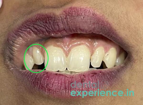
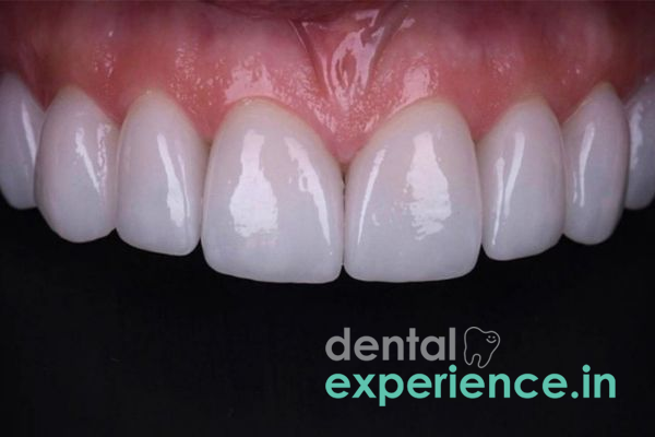
Peg laterals occur when an abnormality develops in the upper lateral incisors (front side teeth), due to inadequate amounts of enamel and dentin being deposited. Instead of growing to a normal flat shape they grow to a conical shape and taper to a point. This is due to a failure in the tooth development stage (morpho-differentiation) to adequately deposit the necessary layers of enamel and dentin. Therefore, although it is not an active oral disease; it may cause significant disturbance to the symmetry of the anterior (front) portion of the smile zone and may prompt many people throughout Kerala to pursue specialized cosmetic dental treatments.
Definition: A peg lateral incisor is a benign genetic and developmental anomaly that results in a small, cone-shaped, upper side front tooth.
Primary cause:Polygenic hereditary traits that affect the early structural formation of individual tooth buds.
Core impact:Cosmetic asymmetry in the smile line; structural gaps (diastemas) between the front teeth.
Key modern fix options:Single-visit biomimetic composite bonding; ultra-thin minimal-prep porcelain veneers; or pre-restorative orthodontic realignment.
Treatment urgency: Elective. Treatment may be pursued at any time during late adolescence or adulthood, provided skeletal growth has stabilized.
Identifying a peg lateral incisor primarily involves identifying specific visual and structural anomalies in the anterior (front) upper arch, rather than pain. The symptoms include:
Pointed shape: The tooth will exhibit a clearly defined, sharp, fanned, or conical crown as opposed to a smooth, straight edge.
Pronounced microdontia: The tooth will be obviously shorter and thinner than its corresponding tooth on the opposite side of the mouth.
Gaps/diastemas: Due to the reduced dimension of the crown, large, dark spaces will exist on each side of the affected tooth.
Asymmetric Smile Line: An irregular arrangement in which the central incisors appear excessively prominent due to the diminished size and structural shortcomings of the adjacent lateral teeth.
A peg lateral incisor is essentially a structural variation and therefore represents no urgent medical concern. However, a consultative evaluation is recommended when a visually apparent difference substantially diminishes an individual’s psychological self-assurance or interpersonal comfort level.
Clinically relevant evaluations are also needed when an undersized tooth leads to related food lodging, thereby causing localized plaque accumulation along the gingival tissues.
Further, when a pointed front tooth genetic trait exists simultaneously with a fully absent tooth on the opposite side, it is advisable to consider an early consultation to avoid potential future changes to the positioning of the entire occlusion.
Genetics are the main underlying factor contributing to an undersized lateral incisor. A slight change or familial hereditary defect makes ameloblasts (the cells forming enamel) produce very limited amounts of external enamel tissue. Since this structural anomaly can be transmitted genetically among family members over several generations, this is a typical inherited facial characteristic found in many families in Kerala.
Although anyone can experience localized microdontia, several factors increase the likelihood of experiencing such a dental variation:
Family History: Having first degree relatives with microdontia or missing front teeth significantly increases the likelihood.
Ectodermal variation: Minor genetic defects impacting skin, hair, and nails may sometimes influence the size and/or shape parameters of the enamel matrix.
Systemic disruptions: Environmental stressors or maternal nutritional differences during pregnancy when tooth buds were forming their original shapes.
Self-diagnosis of a peg lateral incisor can be performed at home with a clear mirror under direct illumination. Simply ask yourself these questions prior to consulting a dentist:
Is the tooth located directly next to my two major front teeth significantly smaller, narrower, or shaped like a small cone or peg?
Are there obvious open spaces/gaps on both sides of this particular tooth, even though my remaining teeth are aligned closely together?
Does my contralateral (opposite) lateral incisor appear perfectly normal, or is it absent entirely?
If you answered “yes” to any of these three questions you are probably viewing a classic peg lateral formation as opposed to a fractured/damaged tooth.
A definitive diagnosis of peg lateral is made promptly upon completion of a thorough clinical examination by a qualified dental practitioner. For accurate planning of treatment options the practitioner will conduct a comprehensive visual assessment along with digital intraoral X-rays (radiographs). Digital X-rays allow assessment of the health and morphology of the underlying root structure. Advanced cosmetic practices in Kerala use 3D intraoral digital scanning technology that allows creation of an interactive digital smile design model. This model illustrates precisely how the dimensions of the tooth correlate with optimal facial proportions.
|
Severity Level |
Structural Presentation |
Orthodontic Impact |
|
Mild |
Mildly narrow crown with normal length; minimal space discrepancy. |
No prior movement needed; direct restoration possible. |
|
Moderate |
Distinctly conical or pointed shape; highly noticeable gaps on both sides. |
Requires minor alignment to distribute gaps evenly. |
|
Severe |
Severely compromised, minute tooth structure; often paired with a missing tooth on the opposite side. |
Requires comprehensive orthodontics before restorative work. |
Restoring a peg lateral has undergone tremendous advances with conservative methods that preserve enamel. Today’s preferred treatment strategies concentrate on enhancing the structural integrity of the tooth versus reducing healthy enamel:
Biomimetic composite bonding for peg laterals: This innovative method consists of sequentially applying thin layers of dental composite resin directly onto the natural surface of the tooth without disrupting any internal structures. The applied material emulates the optical reflective characteristics of natural enamel.
Ultra-thin, custom-fabricated porcelain veneers for small/lack-of-tooth structure: Laboratory-created shell veneers (approximately .3 mm – 0.5 mm thick) are attached to the frontal aspect of the tooth using strong adhesives providing excellent color retention and longevity.
Orthodontics & peg lateral smile design: Prior to enhancing the shape/size of the small tooth, if displaced neighboring teeth have shifted as a result of space loss created by the tooth’s lack of bulk, then clear plastic aligners or conventional braces are initially used to restore ideal spacing relationships prior to enhancing the shape/space requirements of the tooth.
Costs associated with reshaping a small front tooth in Kerala depend on selection criteria including type of material selected, cost associated with clinic location, and extent of complexities involved in restoring shape.
Composite dental bonding: This is an immediately accessible direct approach. Most clinics charge anywhere from ₹2,500 – ₹5,500 for each tooth based on creativity/artistic skills displayed by clinician and quality of composites utilized.
Porcelain veneers/minimal prep Shells: Premium long-term aesthetic alternatives. Costs related to creating lab-based veneers typically fall in the range of ₹10,000 – ₹18,000 for each tooth.
Pre-treatment Orthodontics: If dental arches need to be moved using clear aligners/brackets to establish proper spacing requirements around the microdontic tooth, costs may range from ₹30,000 – ₹1,50,0000 for comprehensive smile makeover plans.
Only experienced professionals in dental aesthetics can successfully reconstruct an ideal-looking version of a peg lateral incisor. Both prosthodontists and those specializing in cosmetic dentistry with extensive experience in creating high-quality aesthetic porcelain/composite reconstructions should ideally take care of reconstructing a peg lateral incisor’s shape/structure. If there are uneven gaps present in areas surrounding the microdontic tooth, an Orthodontist would first correct any misaligned roots to bring them back into aesthetically pleasing alignment.
There is no medically pressing reason to correct a peg lateral incisor. Since it is simply a benign developmental variation, patients may elect to correct this anomaly at their convenience. Recommendations from clinicians suggest waiting until late adolescence or early adulthood (typically post-skeletal maturity age) before placing irreversible treatments such as porcelain veneers on an unduly developed tooth structure. Waiting for skeletal maturity ensures that growth patterns are stable and gum line development has reached its ultimate adult configuration.
Will I be able to get my peg lateral fixed within one day?
Yes! With recent advancements in biomimetic composite bonding; a Cosmetic Dentist may rebuild an undersized pointed tooth in just one session lasting less than one hour!
Is fixing a peg lateral painful?
No! Because modern methods emphasize adding material to existing structures instead of removing material from them; almost all procedures are totally pain-free and non-invasive, usually requiring no anesthesia.
How long will the cosmetic restoration remain viable?
High grade composite bonding remains viable and attractive for approximately five to seven years with regular oral hygiene; whereas premium thin porcelain veneers can survive longer than fifteen years prior to needing repair/maintenance
The old dental practice of aggressively grinding down a small tooth to place a thick crown is obsolete. Modern cosmetic protocols prioritize enamel preservation. By utilizing ultra-thin restorations and biomimetic bonding materials, we can achieve beautiful facial symmetry while protecting the long-term vitality of the tooth structure.














.jpg)
.jpg)

.jpg)
.jpg)

.jpg)
.jpg)
.jpg)
.jpg)
.jpg)
.jpg)
.jpg)
.jpg)