Non-transient, sharp-to-the-point-of-pain sensation in teeth, resulting from friction from toothbrush bristles with an exposed area of dentin; caused by erosion or abrasion of the enamel layer at the cervical neck or cementum layer at the roots and therefore exposing dentinal tubules. This condition is referred to as either a non-carious cervical lesion (NCCL), or dentine hypersensitivity (DH); the dentinal tubules are filled with fluids which can be activated to move by physical (tactile), temperature (thermal) or chemical (osmotic) stimulation, and activate nerves located in the central pulp chamber.
Clinical Definition: Sharp transient pain felt when brushing your teeth due to mechanical irritation of exposed dentin surface by the bristles of your toothbrush.
Red Flags for Immediate Attention: Continuous throbbing, localized gum inflammation/bleeding during light touch/contact, or visible dark grooves (notch) around the gum line.
Intervention Options for Treatment: Application of a desensitizing varnish while you're at our office, placement of resin bonded restorations over the affected area of your tooth, use of soft bristled brushes, and/or gingival grafting.
Sharp Evoked Pain: Nerve twinges which are sudden in nature and have a very short duration caused by the specific stimulation from bristles touching the cervical one-third (the gum line) of the tooth.
Hypersensitivity to Tactile Stimulation: A sensation of pain or discomfort felt while stroking with your fingernail, toothpick, or a stiff-bristled brush over the outer surface of the affected area.
Reaction to Cold Air: Sensations of sharp pain upon breathing in cold air rapidly into the mouth either at the same time as or before brushing.
Visible Evidence of Cervical Wear: Small V-shaped or U-shaped grooves located in close proximity to the juncture of the enamel and the root edge.
Erythema Gingivae: Reddened gums, along with slight recession of gum tissue and exposed darker, yellow-colored dentin surfaces.
Minor sensitivity is indicative of some degree of dentine exposure. Certain signs may indicate significant structural damage; pulp involvement; or destructive activity within the periodontal tissues indicating urgent medical attention:
Pain Without Provocation: Throbbing pain experienced spontaneously and unassociated with any form of mechanical stimulation or thermal activation. This represents an irreversible state of pulpitis.
Sleep Disturbances Due to Pain: Pain that occurs during sleep or intensifies once lying down due to elevated cranial blood flow.
Suppuration/Swelling Locally Associated with the Affected Tooth: Exudate of pus; blistering of gum tissue (parulis); localized swelling on the face near the sensitized tooth.
Persistent Thermal Sensitivity: Duration of cold or heat sensitivity greater than 10–15 seconds post-stimulus.
Tooth Mobility Pathologically Increased: Increased mobility/shift of the affected tooth during both brushing and mastication.
A variety of causes exist for cervical abrasion and erosion:
Mechanical Abrasion: The use of aggressive horizontal brush strokes with a stiff-bristled toothbrush will strip away the protective layer of cementum on all exposed root surfaces.
Cervical Abfraction: This is an abnormal, pathological bending of the tooth structure which can be induced either through heavy biting forces or nocturnal grinding or bruxism; both actions create small cracks in the area where the gum meets the teeth.
Acidic Erosion: Foods and drinks that are consistently in contact with your teeth (i.e., tamarind, raw mangoes, fermented rice water) have low pH levels which dissolve the hydroxyapatite crystal structure allowing the dentinal tubules to open up.
Periodontal Recession: Due to chronic inflammation of the gums and loss of the attachment apparatus, previously hidden areas of sensitive root dentin become visible as periodontal pockets deepen.
Recent Dental Therapies: Patients may experience temporary post-procedure sensitivity after a number of dental treatments including: deep scaling, professional prophylaxis, or chemical bleaching.
Diets High in Acids: Tamarind, raw mangoes, fermented rice water, or drinking black tea with lemon frequently erode enamel.
Habitual Hard Bristle Misuse: There is widespread cultural acceptance of firm toothbrushes or coarse traditional herbal toothpowder misuse that scrape or rub against the thin cervical enamel.
Gastroesophageal Reflux Disease (GERD): When stomach acid regularly flows back into the esophagus it forms a coating on the inside surface of the teeth causing a weakened mineralized enamel.
Gingival Biotype: A patient has inherently thin gums that easily recede when subjected to minor injury.
Untreated Bruxism: Tooth clenching occurs unconsciously and induces high levels of mechanical stress concentration in localized areas around the crown/neck junction of each tooth causing the enamel prism to "pop" off.
You can do an initial, non-aggressive mirror check at home with proper illumination:
Lighting and Visibility: Position yourself in front of a light source. Using your clean fingers, gently pull your upper lip down.
Visual Examination: Look into the mirror and observe for V-shaped grooves on either side of the roots of your teeth where they meet the gum line. You may see black or yellow staining and you may be able to see the recession of the tissue around the root of each tooth.
Initial Touch: Gently run a single bristle of an ultrasmooth toothbrush against the suspected area. Determine if there is a sharp sensation upon contact.
Temperature Test: Swish room temperature water through your mouth. Will pain emanate from one particular tooth or will it come from all four sections of your mouth?
Tissue Evaluation: Do your gums look inflamed? Are they red? If you lightly touch them with a bristle from a soft toothbrush, do they bleed?
To separate the diagnosis of superficial hypersensitivity from deep pulp disease dentists use a systemic approach to diagnose:
Air Jet Test: Dentists are able to isolate which teeth are causing the hypersensitivity and measure their level of hypersensitivity using a series of pulses of compressed air directed to the cervical margins of the teeth.
Exploration: A standardized endodontic explorer (Yeaple) measures how much force is needed to cause the tooth to become sensitive.
Radiographic Examinations: Periapical radiographs taken intraorally provide information about the internal dimensions of the pulp chambers, help eliminate proximate interproximal decay, and determine the health of the supporting bony structures.
Vitality Tests: Endo-ice thermal tests and EPT are used to determine if the nerves within the tooth have died.
|
Grade Level |
Structural Presentation |
Clinical Symptoms |
Typical Interventions |
|
Grade 1: Mild |
Microscopic tubule exposure; no visible tissue loss. |
Fleeting discomfort only with cold air or firm brushing. |
Potassium nitrate paste, varnish application. |
|
Grade 2: Moderate |
Visible V-shaped cervical notch; minor gingival recession. |
Sharp, predictable twinge during every brushing session. |
Glass Ionomer (GIC) or composite resin filling. |
|
Grade 3: Severe |
Deep dentinal loss; active gum recession exposing roots. |
Severe pain during brushing; mild thermal lingering. |
Cervical restoration, gingival grafting, nightguard. |
|
Grade 4: Advanced |
Exposed dentin with pulpal proximity or nerve exposure. |
Spontaneous throbbing, extreme touch and cold pain. |
Endodontic root canal therapy, full crown. |
Restoration treatments have moved toward modernized, low-invasive techniques using biomimic materials to treat exposed dentine as follows:
In-Office Desensitizing Treatment: Applying glutaraldehyde-hydroxyethyl methacrylate (HEMA) or calcium sodium phosphosilicate (bio-glass) pastes onto exposed dentinal tubules will seal them right away.
Glass Ionomer Cement (GIC) Restoration Treatment: The fluoride releasing restoration material binds directly to dentine, fills in the cervical groove, and continues to protect from secondary decay.
Nanohybrid Composite Resin Restoration: Color matched resin matrix is bonded into a cervical defect that restores lost tooth contour and provides excellent aesthetic matching.
Subepithelial Connective Tissue Grafting: Surgical microsurgery by an accredited periodontist covering the root surface(s) that are exposed with soft tissue eliminates sensitivity permanently.
Low Level Laser Therapy (LLLT): Diode laser irradiation changes the membrane potential of nerves and stimulates tertiary dentin formation in the pulp.
|
Procedure |
Average Cost Range (INR / ₹) |
Financial Frameworks & Coverage |
|
Application of Desensitizing Varnish |
₹800 – ₹1,500 per application |
Out-of-pocket payment at all major cities' private dental clinics including Kochi, Trivandrum, and Kozhikode. |
|
Cervical Filling Using GIC / Composite |
₹1,200 – ₹3,000 per tooth |
Out-of-pocket; based upon brand of materials used for restoration and amount of loss present in cervical area. |
|
Periodontal Gum Grafting |
₹8,000 – ₹18,000 per quadrant |
Dependent upon the number of quadrants treated. This procedure must be done by an accredited periodontist. |
Frameworks of Coverage: There are no public health programs that provide coverage for outpatient dental services. Therefore, patients pay out-of-pocket for these services. Patients who receive inpatient surgical admission may be eligible to receive reimbursement through secondary private corporate insurance plans for medically necessary periodontal procedures.
General Dental Practitioner (GDP): Early screening; desensitizing agent application guidance; soft-bristle tooth brushing technique guidance; simple GIC fillings.
Periodontist: When cervical wear is due to advanced chronic periodontitis requiring mucogingival surgery or extensive gum recession or root exposure.
Endodontist: When cervical wear is so deep it has reached the pulp of the tooth resulting in constant throbbing pain, deep infection or requires a root canal treatment.
Prosthodontist: When there are complex cases such as bite misalignment causing abfraction or generalized occlusal wear which will require full mouth reconstruction.
Emergency Care Within 2–4 Hours: Unprovoked throbbing pain; visible swelling of face; facial trauma; unable to swallow due to spreading infection.
Priority Care Within 24–48 Hours: Lingering pain longer than 20 seconds after brushing teeth; bleeding gums; lost cervical filling(s).
Elective Care Within 1–2 Weeks: Twinge pain only triggered by brushing teeth; manageable with desensitizing toothpaste; routine check-ups.
Why am I experiencing pain in my teeth after using warm or cold water while brushing?
Cold water causes rapid contraction and outward movement of fluid within your open dentinal tubules, whereas hot water causes inward displacement. The resulting rapid movement of this liquid will stimulate the nerves beneath your tooth and cause a sudden shooting pain. Desensitizing toothpastes work by sealing the entrances to open dentinal tubules within two to four weeks of continued regular use.
Can natural remedies such as salt water or clove oil help alleviate brushing sensitivity?
Saltwater rinses help soothe inflamed gum tissues, while clove oil contains eugenol, a natural agent that temporarily numbs nerve endings in the tooth. However, neither salt nor clove oil can replace lost enamel structure on your teeth and therefore cannot be used to create a permanent barrier to seal dentinal tubules.
How long does it take for desensitizing toothpaste to begin to work?
Desensitizing toothpastes typically have one active ingredient; either potassium nitrate, which takes approximately seven to fourteen days to build a protective plug at the ends of dentinal tubules and desensitize nerve fibers, or stannous fluoride, also taking approximately seven to fourteen days.
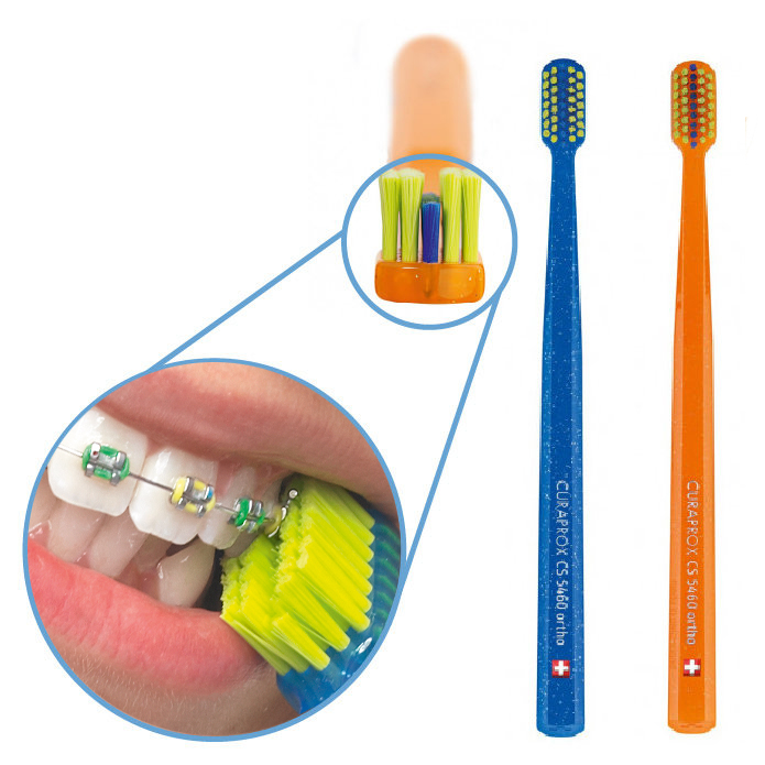
Structural loss is one of the primary causes for mechanical habit (sensitivity) when brushing. Brushing an exposed dentin surface creates a mechanical stimulus that deforms and shifts fluid inside open dentinal tubules, rapidly triggering nerve fibers at the pulpal border. Successful long term management of these patients involves both short and long term strategies. The short-term strategy includes immediately using desensitizing varnish or biomimetic composites to seal the tubules as soon as possible after exposure. The long-term strategy includes changing all patient oral hygiene habits to be non-destructive. These changes include using ultra-soft toothbrushes, eliminating abrasive powders, brushing with circular motions, and controlling any other risk factor contributing to damage (i.e. acid reflux or bruxism).


.jpg)
.jpg)
.jpg)
.jpg)
.jpg)
.jpg)
.jpg)
.jpg)
.jpg)
.jpg)
.jpg)
.jpg)
This site is part of the DEC Dental Network — a Dental Equity & Connectivity ecosystem of clinical, educational, and patient-support brands operated by Team Dental Pvt. Ltd. Our network works as one — guiding you from your first search to your final smile.
© 2026 Team Dental Pvt. Ltd. · Founded 2013, Angamaly, Kochi, Kerala
About DEC Dental