While widespread gaps are categorized as generalized spacing, a localized gap between two adjacent teeth is referred to as a diastema in a clinical sense. A gap between teeth is considered a form of misalignment (malocclusion). Gaps may appear between one or more sets of teeth. Most commonly, spacing appears between the upper front teeth (central maxillary incisors), but can occur anywhere along the dental arches. In terms of function, spacing creates a disruption in the tightly connected, supportive arrangement of teeth in a healthy jaw. Each tooth in a healthy jaw provides lateral support for each of its neighbors. Collectively, they absorb and distribute masticatory loads. When there are gaps between teeth, the individual support provided by each tooth is reduced. Consequently, individual teeth are more susceptible to tipping or shifting under the influence of chewing pressures. In terms of classification in orthodontics, spacing is listed among crowding and rotation as a basic tooth-position problem. It represents a balance between the total size of teeth and the standard dimensions (length/width) of the jaw bone.
Definition: Clinical definition of diastema is the excess gap between the teeth producing a noticeable gap between adjacent teeth.
Warning Signs: Visible spaces with bleeding gums, loose teeth, halitosis (bad breath), or visibly increasing spaces.
Treatment Options: Orthodontic brackets/clear aligners, composite resins/dental bonding, porcelain veneers, and/or frenectomy procedures (surgical excision of excessive labial frenulum).
Consequences of Neglect: Food entrapment, localized periodontitis, bone loss, possible speech disorders, and continued movement of teeth.
Individuals who have spacing will typically identify one or more symptoms:
Gap: The most common symptom associated with spacing is a clearly identifiable gap between adjacent teeth when viewed in a mirror or when smiling.
Entrapped Foods: Fibrous food products such as beef, chicken tend to be wedged painfully into the gap between the teeth.
Gum Sensitivity: The exposed gum tissue (interdental papilla) in the empty space tends to exhibit inflammation and sensitivity.
Speech Alterations: The presence of air passing through the gap may produce a slight whistle/lisp sound, particularly during pronunciations that require the production of "s" or "z" sounds.
Appearance Shift: Gradually expanding appearance of the gap over several months to years indicates that the teeth are continuing to tip further outward.
While some individuals may simply have mild spacing that presents little more than an aesthetic concern, there are several clinical signs that suggest serious problems including damage to deeper structures/tissues or systemic diseases that need immediate attention:
Loose Tooth: If a tooth involved in spacing is mobile (loose/wobbly) upon examination with the tongue, it generally implies significant bone loss has occurred beneath the attached gum tissue.
Progressive Widening: Rapid expansion of gaps observed over several weeks/months usually signifies active destruction of periodontal tissues and/or destructive oral habits.
Bleeding Upon Contact: Bleeding occurs spontaneously from the gum tissue contained within the gap either during routine brushings or independently; it generally indicates extensive pocket formation where bacterial activity is destroying attachment fibers.
Pocket Depth: The sensation that food is being trapped inside a deep periodontal pocket formed below the gum line, accompanied by concurrent offensive odor/halitosis, suggests that bone destruction is actively occurring.
Spacing is primarily influenced by a combination of biological/mechanical behavior and factors:
Genetic Mismatch (Tooth/Jaw): Genetics play a role when patients inherit smaller-sized teeth from one parent and larger jaw bones from another resulting in excess room in the arch.
Large Labial Frenum: A hyperplastic labial frenum (an unusually large or low attaching frenulum) causes the soft tissue connecting the inner surface of the upper lip to the gums to act as a wedge and push the front teeth further apart.
Bone Loss Due to Gum Disease: Chronic gum disease causes destruction of the alveolar bone supporting the roots of teeth. Absence of this solid support structure results in instability in teeth under normal chewing pressure resulting in drifting apart.
Abnormal Tongue Movement: Abnormal swallowing patterns involving pushing the tongue forward against the front teeth rather than toward the roof of the mouth results in additional mechanical stress on the teeth resulting in forward/outward tipping.
Absent/Missing/Peg-Shaped Teeth: Missing/congenitally absent or peg-shaped (small, conically shaped) lateral incisors allow adjacent teeth to migrate into empty spaces.
Certain habits or conditions present in an individual's past medical history increase his/her risk of developing pathological spacing:
Untreated Gum Disease: Individuals with genetically weakened gum attachment mechanisms experience accelerated bone loss resulting in faster tooth migration.
Macroglossia: Large tongues continuously exert involuntary pressure outward against dental arches throughout an individual's lifetime.
Chronic Hard Food Chewing Habits: Dietary consumption of excessively high fiber foods that provide no yielding properties to protect interdental areas from hard food particles may result in increased bone loss as soon as a small gap develops.
Arch Changes Resulting From Age: As humans age, their natural tendency for anterior teeth to gradually drift mesially may become unstable if posterior teeth are lost; this causes a posterior bite collapse, where the increased mechanical force forces the anterior teeth to flare and spread further apart.
Use a clean mirror and sufficient illumination to visually inspect your dental arches for gaps using both vision and touch:
Visual Alignment: View yourself smiling naturally and observe for any spaces where gum tissue surrounding each tooth is entirely exposed rather than protected by close contact between each pair of adjacent teeth.
Floss Test: Use a piece of dental floss to pass vertically downward through the contact point of suspected teeth.There should be a pronounced snap/resistance felt when passing floss through contact points between adjacent teeth. If you find that floss glides easily through without contacting both teeth simultaneously, then there is likely a clinical gap existing between those two adjacent teeth.
Mirror Symmetry: Examine your facial midline carefully. Evaluate if your two upper front teeth are spreading away from each other or if one tooth leans at a greater angle than its counterpart.
Inspecting Soft Tissue Under Lip: Pull up your upper lip gently. Determine if your low-attaching maxillary labial frenulum extends completely down into the empty space between your upper front teeth. This upper tissue attachment turns white/blanches when stretched.
Note: Never attempt to probe or evaluate spaces between teeth using tooth picks/safety pins/metal devices. Probing/evaluating spaces between teeth using metal objects may result in lacerating thin gum tissues and potentially irreversible bone loss.
Each of the above methods must be evaluated individually by trained dental professionals using various diagnostic equipment to establish an accurate diagnosis:
Clinical Evaluation: Using specialized instruments such as periodontal probes, determine exact distances between adjacent teeth and check for unseen gum pockets located within the open spaces.
Radiographic Imaging: Capture intraoral radiographs (periapicals/bitewings) to assess health status of alveolar bone. These radiographs help determine if spacing is merely benign or is a result of bone destruction secondary to chronic gum disease.
Panoramic Radiograph (OPG): A panoramic radiograph shows complete views of both jaws. This enables the dentist to identify congenital missing adult teeth or impacted extra (supernumerary) teeth (mesiodens) inside bony structures that could possibly block normal alignment of anterior/posterior segments of dentition.
Diagnostic Models-Casts: Utilizing intraoral 3-D scanning technology creates precise digital casts/models of dental arches. Dentists utilize these digital models to calculate the millimeters difference between actual tooth mass and jaw length.
Spacing can be categorized clinically into three levels of severity. Each level determines appropriate treatment protocols:
|
Level Of Severity |
Measurement |
Typical Structural Presentation |
Common Treatments Approaches |
|
Minimal |
< 1mm |
Limited small isolated space; almost always limited to central maxillary incisors; no bone loss/tooth mobility |
Cosmetic bonding/composite resins/minimal clear aligners |
|
Intermediate |
> 1mm - < 3mm |
Evident spaces present between multiple teeth; minor tipping of adjacent teeth; initial evidence of trapped food |
Full orthodontic treatment using clear aligners/traditional bracket-based appliances; placement of multiple porcelain veneers. |
|
Extreme |
> 3mm |
Widespread spacing; extreme tooth migration; often accompanied by bone loss/severe tongue thrust habit. |
Combination surgical frenectomy/prolonged orthodontic therapy/permanent fixation. |
Currently available modern treatments offer several alternative restorative/reparative therapies to effectively seal gaps:
Bonding With Composite Resin/Dental Bonding: Mild cases can be treated conservatively with direct application of aesthetically pleasing metal-free composite resins onto sides of affected teeth to widen them slightly closing gaps. This method requires minimal removal of enamel; completed within one visit.
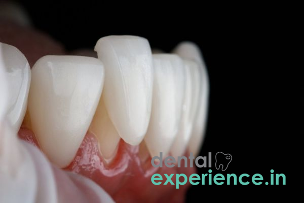
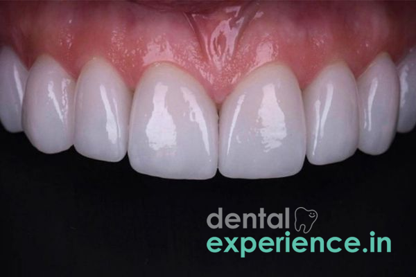
Veneering/Crowning With Porcelain/Ceramics: Cases with spacing plus irregular shape/color defects can be treated using custom fabricated ceramic shells that are cemented permanently onto frontal aspects of affected teeth, creating an even-smooth appearance.
Traditional Orthodontic Brackets/Clear Removable Aligners: Moderate-severe spacing can be corrected using conventional ceramic brackets/removable clear aligners applying constant gentle mechanical stresses to slowly shift each tooth across bone into proper positions/contact points.
Labial Frenectomy Surgery: If an oversized labial frenulum is identified as the main cause for spacing, a minor laser/scalpel surgical procedure can be performed releasing low attaching soft tissue enabling adjacent teeth to move together without being displaced backward again.
Kerala dental costs differ based on severity of gap, choice of material and location (urban hub locations such as Kochi/Thiruvanthpuram vs. rural).
Resin-Bonding: Typically ranges from Rs.1500-Rs.4500 per tooth making it relatively inexpensive cosmetic correction.
Porcelain Veneering/Crowning: Costs for premium ceramics can range from Rs.12000-Rs.25000 per tooth due to laboratory preparation fees.
Orthodontic Treatment: Metal-bracket based treatment can range from Rs.30000-Rs.50000 whereas aesthetics ceramic appliance costs can range from Rs.45000-Rs.70000.
Clear Aligners: High-end Clear Aligner Systems can cost from Rs.60000-Rs.250000 depending on complexity of case
Dental spacing management may require a team effort depending on what caused the space to begin with:
Orthodontist: Responsible for developing and implementing brace and clear aligner therapy plans to actually move teeth.
Prosthodontist/Cosmetic Dentist: The experts who provide the creative add-ons such as custom-designed porcelain veneers, crowns or other forms of full mouth reconstructive cosmetics.
Periodontist: Required if the patient has severe gum disease resulting in excessive space between the teeth. The dentist will need to restore support to the bone beneath the gum line before attempting any movement of the teeth.
Oral Surgeon: Used to perform precise laser or scalpel frenectomies when a significant muscle attachment is blocking closure of the space.
Spacing does not usually present itself as a mid-night emergency situation requiring immediate attention. That being said, there is a defined time frame for how quickly action should be taken relative to when you identify a problem:
Immediate Action (within 24 hours): If one or all of the following occur at once: a tooth which was previously spaced becomes loose; an acute pain develops in one or both of your spaced teeth; trauma occurs to a spaced tooth due to a hard food object causing a laceration to form in the soft gum tissue protecting the tooth.
Elective (within 1 – 2 months): Generally recommended for standard developmentally-caused, genetically-caused spaces. Scheduling an appointment prior to having the teeth professionally aligned allows for the prevention of continued tipping of adjacent teeth into the space and helps protect the interdental bone from years of erosion through constant food debris.
Are adults capable of experiencing new gaps opening up between their teeth if their teeth were completely straight as children?
Absolutely. This is a very common occurrence among adults and it generally results from a prolonged loss of the underlying bone that supports the teeth. Most commonly this is caused by some form of chronic periodontal disease, but can also result from chronic habit formation (i.e., tongue thrusting).
How painful is a surgical frenectomy, and do I have to undergo surgery before I get braces?
A frenectomy is an extremely minor, quick outpatient procedure that is performed under local anesthesia. When using modern dental lasers to execute a frenectomy, virtually no discomfort is felt during or immediately following the procedure. A frenectomy is only required if a thick band of tissue presents a high risk of pushing the teeth back apart. Clinically, orthodontic appliances are used to bring the teeth together first, and the surgical frenectomy is performed afterward to prevent the space from reopening.
Will my newly closed spaces remain closed after my orthodontic treatment is complete?
Your teeth naturally want to go back to where they were before, so unless you wear a fixed or removable retainer after treatment, they will likely continue to spread apart over time. In order to maintain proper alignment and prevent future gaps, it is essential to use an appliance (such as a fixed lingual retainer) to keep your front teeth locked together for long-term retention, which requires life-long dental monitoring.
The ultimate goal in achieving clinical success in correcting dental spacing depends solely on determining the appropriate reason why spacing exists prior to providing treatment. Correctly completing cosmetic bonding or using aligners without initially addressing active periodontitis or accurately diagnosing a hyperplastic frenum will ultimately lead to long-term failure. Achieving a stable result necessitates creating an ideal relationship between alveolar bone support, balanced musculature forces from the tongue and lips, and rigid compliance to retention protocol guidelines after treatment completion. Patients must understand that closing a space is not merely an aesthetically pleasing adjustment, but rather, it represents a critical structural adjustment that provides protection to the combined health of the entire dentition.














.jpg)
.jpg)

.jpg)
.jpg)

.jpg)
.jpg)
.jpg)
.jpg)
.jpg)
.jpg)
.jpg)
.jpg)