Dental discoloration or staining occurs when pigmented substances accumulate on the surface of a tooth as chromogenic substances on the enamel matrix or absorb into dentinal tubules through internal discoloration. The classification for dental discoloration within restorative dentistry is as follows: Extrinsically stained teeth have surface level deposits onto the pellicle; while intrinsically stained teeth exhibit deep structural discoloration within the hydroxyapatite crystal lattice. If not treated, an extrinsically stained tooth will continue to develop and potentially form plaque biofilms which could lead to further development of the tooth enamel's demineralization and eventual progression towards tooth decay.
Clinical Definition: Discoloration caused by chromogens, lifestyle choices, or cellular changes in dentin that appear as either surface or structural discolorations.
Critical Indicators of Urgent Care: A dark black spot lesion close to the gum line indicating possible gingival recession, sudden lone tooth darkening suggesting necrosis of the tooth pulp, or extreme tooth sensitivity upon exposure to cold/hot temperatures at the time of developing a stain.
Recovery Options: Laser/LED whitening in office, microabrasion, porcelain veneers, and professional prophylaxis (scaling and polishing).
Regional Cost Average: Depending on the type of procedure needed (i.e., simple scaling & polishing vs. advanced cosmetic restoration), costs range from ₹800 to ₹35,000+ in clinics located in Kerala
Color Shifting: Yellow bands, brown lines, or chalky white spots (fluorosis) appear on the anterior teeth.
Lack of Luster: The enamel loses its natural shine and appears dull or opaque in lighted conditions.
Texture Changes: The high deposits (most commonly behind lower incisors) will be rough and have a "grain" that can be felt by the tongue.
Single Tooth Shadowing: One tooth becomes gray, blue, or dark brown after previous traumatic injury to the tooth or after endodontic therapy (root canal).
The majority of all tooth discolorations are merely cosmetic; however, there are several symptoms that may point toward serious dental disease or pathologies affecting the deeper tissues:
One-Sided Staining: If one tooth has changed from normal color to grayish-purple, this could be an indication of necrotic pulp (the nerve tissue within the pulp chamber is dead).
Pulsating Pain: Pulsating pain while eating or drinking warm/cold foods indicates that you have deep caries lesions in your teeth.
Swollen Gingiva: Teeth with significant discoloration along with swelling of the gum surrounding the tooth, which can result in bleeding or pus production, indicate a periodontal abscess or advanced infection.
Consumption of Chromogenic Beverages: Long-term frequent consumption of beverages containing high levels of tannins (such as black tea [chaaya], filter coffee, karkidaka kanji herbal infusion, or pathimugam herbal water).
Consumption of Deep Colorful Culinary Spices: Increased use of spices with high amounts of color such as turmeric (manjal), red chili powder, and heavy tamarind-based gravies which have penetrated through the enamel over years of time.
Use of Tobacco Products: Deposits of nicotine and tar from using tobacco products (cigarettes or chewing betel leaf [paan] and tobacco).
Dental Trauma / Pulpal Hemorrhage: Physical trauma to a tooth resulting in internal hemorrhaging within the microscopic dentinal tubules, turning the tooth grey over many years.
Antibiotic Use by Children During Enamel Formation: Administration of tetracycline or doxycycline to children early in life when they were still developing their teeth.
Enamel Hypoplasia: Thin or poorly mineralized enamel that is highly susceptible to absorbing darkly colored substances.
Dry Mouth / Xerostomia: Reduced flow rate of salivary secretion can result in a reduced ability of the natural cleaning/protective actions of the oral cavity, decreasing the amount of time it takes for chromogen to bind to the enamel pellicle.
Attrition Due to Age: Over years, attrition occurs resulting in the gradual thinning of the outer transparent enamel layer, exposing the yellowish-colored dentin below.
Inadequate Oral Hygiene / Poor Brushing Habits: Failure to consistently brush your teeth results in sticky bacteria forming a hard deposit known as dental calculus/tartar that is dark, porous, and readily binds to chromogens.
Perform a preliminary inspection prior to a dentist visit in front of a well-lit mirror. The self-exam should include the following steps:
Rinse Thoroughly: Swish plain water around the mouth to remove loose food particles from teeth.
Visual Examination: Inspect teeth under bright, white light. Determine if staining is uniform on all teeth or isolated to one tooth.
Tactile Check: Use a clean fingernail or your tongue to run along the stained portion of the tooth. If you feel an elevation or roughness, then it is most likely to be due to extrinsic tartar/plaque buildup.
Lighting Angle: Point a flashlight at the back side of your upper and lower front teeth. Any tooth that blocks light from entering as compared to its neighbors which are translucent will probably indicate nerve death.
Important Safety Warning: Do not use metal instruments or abrasive products to scrape away stains. This could permanently damage the surface of your tooth.
Dentist’s evaluation of discolored teeth includes both visual inspections, examinations with optical devices, and diagnostic equipment:
Transillumination & Color Matching: Evaluation by means of special optical guide shades and fiber optic light for determining depth and location of intrinsic vs. extrinsic discoloration.
Digital Radiography (X-Ray): Intraoral periapical X-rays to evaluate possible internal resorption of roots, or proximal caries (cavities) which exist below the surface causing discolored teeth.
Pulp Vitality Testing: Electric pulp tester (EPT) or cold spray thermal testing to evaluate if a darkened tooth has live nerve tissue.
|
Grade Level |
Structural Impact |
Clinical Characteristics |
Primary Intervention |
|
Grade 1: Superficial Extrinsic |
Pellicle surface deposit |
Light yellowing; localized tea/coffee stains |
Prophylaxis (Scaling & Air Polishing) |
|
Grade 2: Deep Extrinsic |
Hardened calculus/enamel pits |
Brown/black lines near gumline; tobacco stains |
Ultrasonic Scaling + Air Polishing |
|
Grade 3: Moderate Intrinsic |
Outer dentin layer involved |
Generalized yellow/brown hue; mild aging stain |
In-Office Chemical Bleaching (Carbamide/Hydrogen Peroxide) |
|
Grade 4: Severe Intrinsic |
Deep dentinal matrix damage |
Dark gray/blue bands, tetracycline lines, dead nerve |
Root Canal + Internal Bleaching or Porcelain Veneers |
Modern dental practices are designed to be minimally invasive and to preserve as much of your natural enamel as possible:
Air Polishing & Ultrasonic Scaling: High-frequency sound waves and small amounts of either glycine or sodium bicarbonate powder remove tough stains caused by coffee, tea, and tobacco, all while preserving your enamel.
In-Office Laser / LED Chemical Bleaching: The application of concentrated hydrogen peroxide gel (25–40%), which is then activated by light at a specific wavelength to break double bonds that cause deep organic stains in 45–60 minutes.
Enamel Microabrasion: A microscopic process used to remove surface enamel defects by utilizing a compound made from hydrochloric acid and pumice. This is the best way to treat fluorosis-related white spots located locally.
Internal Non-Vital Bleaching: The walking bleach technique using sodium perborate that is placed inside the pulp chamber of a tooth that has had a root canal performed and is currently dark.
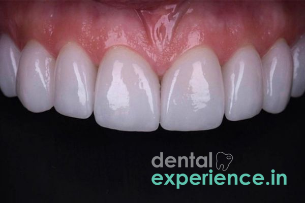
Porcelain Veneers & Composite Bonding: Thin, custom-made porcelain veneers (0.3–0.5 mm) that bond onto the front surfaces of teeth when intrinsic staining cannot be removed through chemical bleaching.
Prices vary among dental clinics throughout Kerala (Kochi to Thiruvananthapuram), depending upon what materials you choose and how extensive the diagnosis is:
Professional Scaling & Polishing: ₹800 – ₹2,500
Each Session of In-Office Teeth Bleaching: ₹8,000 – ₹18,000
Each Enamel Microabrasion Procedure: ₹1,500 – ₹3,500
Each Composite Direct Veneer Procedure: ₹2,500 – ₹5,000
Each E-Max Porcelain Veneer Procedure: ₹10,000 – ₹22,000
Financial Note: Cosmetic tooth whitening is generally considered an out-of-pocket elective service. However, structural restorative services such as root canals or crowns for traumatized discolored teeth may qualify for reimbursement under private health insurance policies or programs if they were deemed medically necessary because of infection or decay.
General Dental Surgeons: They treat basic prophylactic scaling, air polishing, and simple, mild chemical whitening.
Prosthodontists: They are recommended for advanced cosmetic rebuilding of teeth with porcelain veneers, complete coverage ceramic crowns, etc., or full-mouth aesthetic smile design.
Endodontists: They should be consulted if you have darkened a single tooth due to it being non-vital and/or traumatized, requiring an internal bleach or root canal.
Pediatric Dentists (Pedodontists): They treat developmentally formed enamel defects and fluorosis as well as stains on primary/mixed dentition of your child.
Immediate (Within 24 – 48 hours): Throbbing pain or severe facial swelling accompanying the sudden loss of whiteness on one tooth.
Scheduled (1 – 2 weeks): One or two dark brown/black spots near the gum line which feel rough or catch food.
Elective (Flexible): A general feeling of yellowish color throughout your teeth.
How can I have stained teeth when I am brushing with a regular toothbrush at least two times per day?
You are removing all of the loose and soft plaque building up around your teeth. The microscopic pores in your teeth's enamel will still absorb many color pigments from foods you eat every day (like black tea, turmeric, and coffee). In addition to this, hardened minerals of calculus build up on your teeth that will not come off with normal toothbrush bristles and require an ultrasonic tool for a dentist to clean.
Will using charcoal powder or baking soda help whiten my discolored teeth?
No. Abrasive charcoal powder and the harshness of baking soda strip away the protective outer layer of your teeth (enamel). Although it removes the top layer of debris, repeatedly doing so strips away some of the enamel, which exposes the yellowish-colored dentin underneath, causing teeth to appear darker over time.
Are whitening treatments done at the office permanent?
Generally speaking, most whitening treatment effects last between 1 to 3 years. Your diet, how often you consume chromogenic foods (such as dark curry gravies or teas), your smoking habits, and your oral hygiene regimen will greatly determine how long your results last.
To effectively manage tooth discoloration over the long term, you have to identify whether a patient has external staining (pellicle) or internal staining (dentinal). Most modern protocols first try to use non-invasive treatments such as ultrasonic scaling to remove stains from the tooth surface and/or light-activated peroxides prior to doing irreversible procedures that require removal of tooth structure. Post-procedure maintenance is key in maintaining successful treatment results by utilizing non-abrasive fluoride toothpastes after dental treatment, avoiding prolonged exposure of teeth to dark food/drink colors, and having regular scheduled biannual professional cleanings performed so that hardening calcium-based deposits do not damage your enamel.



.jpg)
.jpg)
.jpg)
.jpg)
.jpg)
.jpg)
.jpg)
.jpg)
.jpg)
.jpg)
.jpg)
.jpg)
This site is part of the DEC Dental Network — a Dental Equity & Connectivity ecosystem of clinical, educational, and patient-support brands operated by Team Dental Pvt. Ltd. Our network works as one — guiding you from your first search to your final smile.
© 2026 Team Dental Pvt. Ltd. · Founded 2013, Angamaly, Kochi, Kerala
About DEC Dental.jpg)