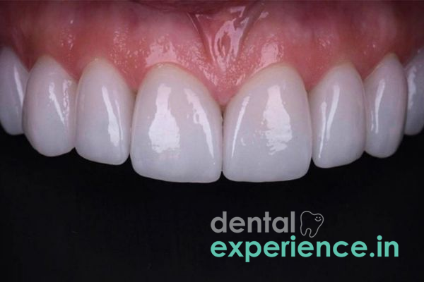
The inherent dental staining phenomenon known as tetracycline discoloration results when a patient receives tetracycline-class antibiotics at any point during active tooth development from birth to approximately 8 years of age or during gestation. Tetracyclines are taken into developing teeth through the bloodstream via ingestion by either pregnant mothers (second or third trimester) or children (from birth to approximately 8 years old). The tetracycline molecule binds to calcium ion sites in developing dentin that have yet to undergo calcification, becoming a permanent component of the crystalline hydroxyapatite structure of the developing dentin as tetracycline-calcium phosphate. Upon eruption of the tooth and subsequent exposure to ambient light, photo-oxidative degradation of the incorporated tetracycline occurs, darkening the initial yellow-brown hues into characteristic grey, blue, or slate bands across the tooth structure. Tetracycline discoloration is classified as a non-carious intrinsic enamel and dentin defect and requires unique cosmetic and restorative treatment modalities.
Clinical Definition: An intrinsic discoloration of the dentin layer created by chemical bonding between an antibiotic (tetracycline) and calcium during tooth formation (birth – 8 years or pregnancy).
Indications of Urgency: Coexistent enamel hypoplasia (reduction/thinning), excessive thermal hypersensitivity, surface microfractures, or anxiety-induced social impairment.
Solutions: In-office deep bleaching (mild/moderate), minimally invasive porcelain veneering, adhesive resin bonding, or complete crown coverage using all-ceramic materials.
Horizontal Chromatic Banding: Alternating bands of grey, brown, blue-violet, or yellow run horizontally across teeth from one proximal edge to the other.
Fluorescent Properties: When viewed under specialized UV light, freshly erupted tetracycline-stained teeth fluoresce brightly in a yellow-green color. Over time, photo-oxidation darkens the hue into dark slate or grey shades.
Opaque Optic Appearance: Stained teeth appear duller than unaffected teeth and lose their natural translucency, displaying a flat, opaque optical appearance originating within the affected dentin layer.
Surface Enamel Roughness: Severe cases (Grade IV) exhibit surface pitting, structural ridges, and hypoplastic enamel development defects.
Mild tetracycline stains are primarily cosmetic issues; however, certain symptoms prompt immediate clinical evaluation:
Severe Enamel Hypoplasia: Defects that expose underlying dentin create an environment prone to rapid caries progression or localized structural collapse.
Thermal Sensitivity: Sudden sharp pain from hot or cold exposure indicates significant enamel thinning or exposed dentinal tubules.
Bleeding & Tissue Recession Around Older Restorations: Gingival recession or bleeding upon probing around existing crowns/veneers can signal microleakage or compromised marginal integrity.
Pathologic Attrition: Excessive or abnormal wear on opposing teeth due to structural compromises in the enamel matrix.
Childhood Antibiotic Administration: Exposure to classical tetracyclines (such as tetracycline or minocycline) in children under 8 years of age during active dentinogenesis.
Maternal Ingestion During Pregnancy: Tetracycline intake by a mother during the second or third trimester affecting the developing primary dentition of the fetus.
Adult Minocycline Use: Long-term adult use of minocycline (commonly prescribed for chronic acne) causing intrinsic discoloration in fully formed teeth through oxidation and dentin matrix binding.
Vulnerability by Age: Children under 8 years face the greatest vulnerability due to active dentinogenesis.
Duration & Cumulative Dose: Prolonged courses or higher doses lead to greater stain deposition and deeper penetration into the dentinal matrix.
Extrinsic Superimposition (Regional Dietary Factors): Regular consumption of darkly pigmented regional dietary items in Kerala (such as black tea [kattan chaya], turmeric-rich fish curries, and betel nut [paan]) adds heavy extrinsic staining over intrinsic bands.
Acidic Enamel Erosion: Frequent consumption of acidic local ingredients (such as kudampuli or tamarind) causes enamel loss, increasing the optical exposure of underlying discolored dentin.
Perform a preliminary self-assessment at home using these guidelines:
Lighting Setting: Use natural daylight or a bright white light source. Avoid yellow household lighting as it distorts natural tooth shades.
Inspect Your Teeth: Pull your lips back to observe your front teeth. Look for distinct horizontal bands running across the teeth (vertical lines generally indicate other conditions).
Identify Color Shade: Note the color spectrum. Stains range from light yellow-tan to dark grey, blue, and purple. Darker shades indicate deeper dentinal involvement.
Check Surface Texture: Pass your tongue gently over the tooth surfaces to feel for smooth versus rough or pitted areas.
Important Safety Warning: Do not use sharp metal objects, needles, or abrasive household products to scrape your teeth.
Visual & Spectrophotometer Analysis: Utilizing a digital intraoral spectrophotometer and standardized shade guides to analyze light reflection and determine stain depth within the dentin.
Medical History Review: Documenting childhood or maternal antibiotic administration history.
Radiographic Evaluation: Intraoral Periapical (IOPA) and Panoramic Radiographs assess pulp chamber dimensions, dentin thickness, root anatomy, and rule out internal resorption.
Pulp Vitality Testing: Electric Pulp Testing (EPT) and thermal cold testing confirm pulp vitality before initiating cosmetic or restorative care.
|
Severity Grade |
Clinical Features |
Structural Depth |
|
Grade I (Mild) |
Uniform light yellow, tan, or grey discoloration across the crown without distinct banding. |
Superficial outer dentin. |
|
Grade II (Moderate) |
Darker, uniform yellow-brown or slate-grey stain without sharp, dark horizontal lines. |
Mid-depth dentin layer. |
|
Grade III (Severe) |
Prominent, dark grey or blue-purple horizontal banding across multiple teeth; highly noticeable. |
Deep dentin tissue close to pulp. |
|
Grade IV (Intense) |
Extremely dark, opaque purplish-black banding combined with structural enamel pitting/hypoplasia. |
Full-thickness dentin & enamel defects. |
Extended In-Office Bleaching: Application of high-concentration carbamide or hydrogen peroxide gels over an extended multi-month protocol; primary utility for Grade I and select Grade II cases.
Ultra-Thin Porcelain Veneers: Custom-crafted ceramic shells (0.3–0.5 mm) bonded to the prepared labial enamel. They provide superior opacity, shade-blocking, and long-term color stability for Grade II and Grade III stains.
Composite Direct Bonding: Layering high-density, shade-matched composite resin directly onto prepared enamel; a conservative, single-visit option for mild to moderate localized staining.
Zirconia or Lithium Disilicate Crowns: Full-coverage ceramic crowns indicated for Grade IV discoloration or severe structural enamel loss to completely mask underlying dark dentin.
The cost of treatment in Kerala varies based on clinical complexity, materials selected, and facility expertise:
Full-Arch In-Office Bleaching (Upper or Lower): ₹8,000 – ₹18,000
Direct Composite Bonding: ₹2,500 – ₹5,500 per tooth
Porcelain / E-Max Veneers: ₹12,000 – ₹25,000 per tooth
High-Translucency Zirconia Crowns: ₹10,000 – ₹22,000 per crown
General Dental Practitioners: Ideal for initial oral health evaluations, routine scaling, preliminary diagnostics, and simple external whitening.
Prosthodontists: Recommended specialists for Grade II through Grade IV cases requiring complex shade-blocking, porcelain veneers, CAD/CAM crowns, or full-mouth aesthetic rehabilitation.
Cosmetic / Restorative Dentists: Specialists trained in advanced composite layering, smile design, and clinical whitening protocols.
Endodontists: Consulted when internal non-vital bleaching ("walking bleach") is required for individual non-vital discolored teeth.
Immediate (Within 24 – 48 Hours): Warranted only if structural enamel chipping creates sharp edges causing soft-tissue trauma or if acute hypersensitivity develops.
Scheduled Consultation (Standard Window): Standard time frame for cosmetic evaluations, diagnostic wax-ups, shade mapping, and treatment planning.
What is the best way to treat tetracycline-stained teeth that have permanently browned in Kerala?
Porcelain veneers or E-Max ceramic crowns represent the most durable, long-lasting cosmetic solutions for masking dark tetracycline banding. While lighter yellow stains may respond to extended bleaching, deep grayish-purple bands are best covered with custom ceramic restorations fabricated by a skilled prosthodontist.
Will chemical tooth whitening be able to completely eliminate deep, dark gray tetracycline bands?
No. Chemical bleaching oxidizes organic stains in the enamel, but once tetracycline has bound to calcium within the dentin matrix, it cannot be entirely removed chemically. Bleaching may lighten the base shade, but custom veneers or crowns are generally required to fully mask dark horizontal bands.
How long will porcelain veneers last when used to cover severely discolored teeth?
High-quality porcelain or lithium disilicate veneers typically last 10 to 15 years with proper oral hygiene and maintenance. Patients should avoid biting down directly on hard substances (such as crab shells or hard candy) to prevent ceramic chipping.
Restoring teeth affected by tetracycline discoloration requires a balanced approach combining high-level aesthetics with maximal tooth structure preservation. Minimally invasive enamel preparation—removing only what is necessary to establish proper bonding and adequate ceramic depth—prevents unnecessary pulpal irritation. Utilizing modern opaque-core ceramics (such as lithium disilicate or layered zirconia) allows clinicians to achieve natural translucency while blocking dark underlying dentinal bands. Long-term success relies on thorough pre-operative shade diagnostics, precise marginal fit, and disciplined post-restorative maintenance.



.jpg)
.jpg)

.jpg)
.jpg)
.jpg)
.jpg)
.jpg)
.jpg)
.jpg)
.jpg)
.jpg)
.jpg)
This site is part of the DEC Dental Network — a Dental Equity & Connectivity ecosystem of clinical, educational, and patient-support brands operated by Team Dental Pvt. Ltd. Our network works as one — guiding you from your first search to your final smile.
© 2026 Team Dental Pvt. Ltd. · Founded 2013, Angamaly, Kochi, Kerala
About DEC Dental