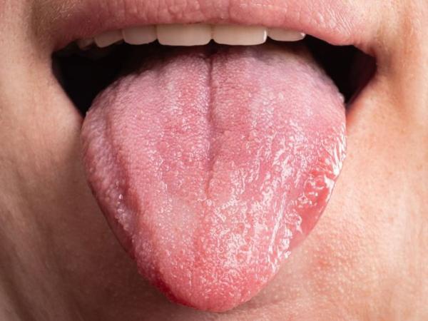
Tongue problems include a wide variety of different types of mucosal, muscle, and blood vessel disorders. The tongue is the most important muscular organ of our body that is protected with a special type of mucosa. This mucosa contains papillae and taste buds. The main functions of the tongue are eating (mastication), swallowing (deglutition), speaking (speech), and tasting (gustatory sensation). These conditions can vary from simple, non-serious inflammatory changes such as geographic tongue (also called benign migratory glossitis) to severe changes in the tongue's mucosa, including black hairy tongue (lingua villosa nigra) and ulcerative forms of candidiasis, leukoplakia, and even oral squamous cell carcinoma (OSCC). In clinical oral medicine, tongue lesions often represent both local pathology as well as various systemic dermatologic, nutritional, and autoimmune diseases.
Clinical Description: Any pathological alterations to the surface characteristics of the mucosa of the tongue, including color, vascularity, and/or tissue structure.
Emergency Indications: An indurated unilateral lesion on the tongue lasting longer than 14 days; angioedema characterized by acute tissue swelling; dysphagia, characterized by an inability to swallow; or severe, painful sensations on the tongue that do not subside.
Major Treatment Options: Antimycotic therapy administered either topically or systemically, nutritional supplements, surgical removal of lesions, laser ablation, and specific oral hygiene techniques.
Surface Appearance Changes: Patches on the surface of the tongue that are white (coated) in appearance; smooth, red surface areas as if they have been burned by fire (redness from atrophic glossitis); wide, deep furrows in the anatomy of the tongue; or blackened papillae (hyperkeratosis).
Pain/Tactile Response: A painful, burning sensation when eating hot, spicy, or acidic foods; sensitivity during mastication; or a lack of a normal sensory response to touch.
Functional Abnormalities: Distorted perception of taste (dysgeusia), reduced mobility of the tongue, or localized swelling resulting in distorted speech patterns (slurring).
Long-Term Hardening of the Tongue: A hard, chronic, sore area that is indurated with an elevated border and has remained so for over 2 weeks; this condition requires a biopsy for potential malignancy.
Airway Obstruction: Rapid onset of severe lingual edema causing upper airway obstruction and/or inspiratory stridor; and/or an inability to swallow saliva due to acute dysphagia/odynophagia.
Fixation: Inability to project the tongue forward from the floor of the mouth (i.e., fixation of the tongue).
Overgrowth of Microorganisms: The overgrowth of Candida albicans is caused by an imbalance in the normal oral flora or localized immunosuppression.
Trauma (Physical/Thermal): Irritation to a tooth or teeth from a poorly placed molar, a fractured dental restoration, an improperly fitting denture, etc., through either mechanical action, thermal action, or both, as a result of an accident while eating.
Dietary/Lifestyle Causes: Frequent intake of foods that are too hot, too spicy, or too acidic (as found in many traditional meals) and/or frequent use of tobacco and/or alcohol.
Nutritional Deficiencies: A prolonged lack of iron, vitamin B12, folic acid, zinc, etc., in the body, which can cause damage to the mucosa in the oral cavity.
Systemic Conditions: Untreated diabetes mellitus, lichen planus, autoimmune diseases, or xerostomia (dry mouth).
Recurring Habits: Chewing betel quid/areca nut (pan masala), smoking, or chronic alcohol consumption.
Visual Inspection: Stand directly in front of a well-lit mirror and open your mouth wide enough to extend your full tongue. Check for abnormal discolorations or abnormal areas of thickening on the top surface of your tongue.
Lateral and Ventral Examination: Use a long, clean piece of gauze to hold the tip of your tongue. Gently pull the tip of your tongue to the left and then to the right. This will allow you to inspect the sides as well as the underside of your tongue.
Palpation of the Tongue: Thoroughly wash your hands. Place your index finger on the top of your tongue and your thumb underneath it. Gently press your fingers together, moving along the tongue to check for any abnormal masses, lumps, or hidden tenderness throughout its length.
Inspection with Lighting: Direct visual inspection utilizing an extremely bright light source and a mouth mirror to visualize mucosal color, texture, and any lesion margins or boundaries.
Palpation: A bimanual evaluation to assess tissue flexibility (the amount of give), the extent to which soft tissue has become hard (indurated), and whether any tenderness is present.
Diagnostic Testing/Biopsy: Excisional or incisional tissue biopsy to obtain a specimen for histological examination, mucosal swabbing to obtain a specimen for fungal culture, or routine blood panels to rule out systemic deficiencies.
|
Severity Level |
Clinical Presentation |
Primary Implication |
Recommended Pathway |
|
Mild / Superficial |
Transient red/white patches, minor aphthous sores, slight papillary change |
Temporary local discomfort |
Conservative hygiene & OTC care |
|
Moderate |
Deep anatomical fissures, widespread candidiasis, painful burning glossitis |
Functional interference, systemic links |
Clinical evaluation & prescription care |
|
Severe / Critical |
Non-healing indurated ulcers, leukoplakia, rapid swelling, tissue necrosis |
Potential malignancy or airway risk |
Immediate specialist biopsy & emergency care |
Pharmacological Treatments: Topical or systemic antifungal medications (e.g., fluconazole) for fungal infections, topical steroid gels for inflammation, and antiseptic mouth rinses containing chlorhexidine gluconate to control bacterial infection and manage plaque buildup.
Surgical and Laser Therapies: CO2 laser ablation or scalpel excision to remove leukoplakia, giant cell lesions, or significant fibrous hyperplasia.
Nutritional and Dental Interventions: High-dose oral or parenteral vitamin supplementation to resolve mucosal deficits, alongside dental restorations (such as smoothing or replacing sharp, damaged teeth) to prevent ongoing mechanical trauma.
Specialist Consultations: Examination by a specialist will cost anywhere from ₹500 to ₹1,500 in most outpatient clinics.
Biopsy Costs: The costs of diagnostic brush biopsy and punch biopsies are generally ₹2,000 to ₹6,000 based upon pathology lab charges.
Hospital Costs: Hospital based laser therapy or surgery for treatment of leukoplakia, and minor surgical excision (e.g. removing fibrous hyperplasia) can range from ₹8,000 up to greater than ₹35,000 out of pocket.
Specialists in Oral Medicine & Oral Pathology: Expertise for identifying the underlying causes for the many different types of systemic mucosal manifestations such as chronic glossitis, and complex premalignant lesions.
Maxillofacial Surgeon: The surgeon is required to perform an invasive excisional biopsy on a mass that could be either benign or malignant, to remove both benign and malignant masses from the oral cavity, and to treat deep tissue trauma.
Dental General Practitioner: Suitable for monitoring all patients regularly, to screen all new patients for abnormalities, to eliminate any mechanical source of irritation to the oral cavity, and to perform all basic oral prophylaxis.
Emergency (Within 2–4 Hours): Swelling of the tongue rapidly progressing to the point where it may cause airway constriction, severe bleeding resulting from trauma to deep structures of the oral cavity, or sudden allergic angioedema.
Urgent (24–48 Hours): Extensive, painful ulcers that are so large they interfere with taking fluids by mouth, or very severe burning sensations that prevent normal nutrition.
Routine/Elective (1–2 Weeks): All asymptomatic white or red patches, persistent smooth surface changes, or routine evaluations of structural fissures.
My tongue burns or feels like fire after I eat something too spicy; why?
The burning usually occurs due to thinning or an inflammatory response of the papillae on the surface of your tongue. The papillae protect your nerve endings. When they are gone, you have exposed nerve endings and they react sharply to the acidic and spicy chemicals in food, creating a "burning" sensation.
Why do I have a thick, white coat on my tongue?
There could be several reasons for having a thick, white coat on your tongue. If the white coat cannot be wiped or scraped away with brushing, it may be a sign of oral leukoplakia or hyperkeratosis. If you can scrape off the white coat and there is redness underneath that hurts when touched, then it's probably a thrush (Oral Candida).
Do the deep cracks or groves on the top of my tongue pose a danger?
Usually, a deep groove in the middle of the tongue represents a non-cancerous structural anomaly called lingua plicata (fissured tongue) . These deep cracks are common places for bacteria and food particles to settle. If not removed by gentle cleaning while you’re brushing your teeth, these areas can become inflamed or develop fungal infections.
The diagnosis of pathologies of the tongue is a methodical process, as there are multiple possible reasons why an individual may have a pathology in this area. Local trauma, oral disease, and systemic conditions all intertwine, making it difficult to determine which factor caused the pathology. Therefore, treatment focuses on finding the underlying cause rather than just treating the symptoms.
If a patient has a persistent, non-healing mucosal alteration or unexplained lingual pain, they need a complete examination performed by a dentist, periodontist, or oral surgeon. This includes, but is not limited to, palpation and biopsy when indicated. Maintaining good oral hygiene, nutritionally supporting healthy mucosa, removing chronic trauma (both physical and chemical), and avoiding common mucosal irritating agents are crucial to long-term mucosal health. In addition, regular evaluations are necessary for early detection and management of potentially malignant lingual lesions.














.jpg)
.jpg)

.jpg)
.jpg)

.jpg)
.jpg)
.jpg)
.jpg)
.jpg)
.jpg)
.jpg)
.jpg)