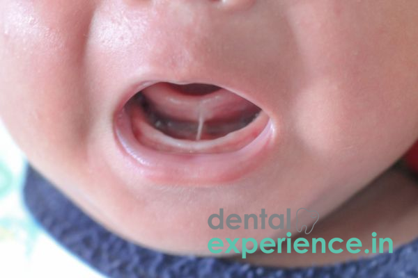
The clinical term for what we commonly refer to as "tongue-tie" is ankyloglossia, which is a common congenital deformity that can affect normal function in the mouth. The deformity occurs when there is a too short, too thin, or too tight piece of mucosa (mucous membrane) at the bottom of the tongue called the lingual frenulum. When this piece of mucosa connects to the bottom of your mouth with an abnormal attachment point, it limits how far forward you are able to move the front part of your tongue. Because the movement of the front part of your tongue affects many basic oral functions, such as suckling/breastfeeding, swallowing, speaking, and maintaining good oral hygiene, the limitation caused by this condition can lead to problems in these areas. As a soft tissue structural disorder, it fits into the category of dental anomalies found within both children and adults. If left untreated, ankyloglossia will cause changes in the way that your mouth works; because of this, other problems may develop in addition to those described above. These additional problems include: dental crowding, narrow palatal arches, and aerophagia.
Clinical Definition: A congenitally restricted ability of the tongue to move due to an abnormally short lingual frenulum.
Urgency: Failure-to-thrive infant, extreme damage to a mother's nipple while breastfeeding, poor articulation in speech, and open-mouth resting position.
Surgical Treatment Options: Laser frenectomy, surgical frenotomy, and post-procedure myofunctional therapy.
Impact: Ankyloglossia is commonly identified among neonates and young children in regional clinical practices—typically during early nursing experience or the first year of schooling.
Tongue Elevated to a Low Degree: The inability of an individual to place their tongue high in the mouth, above the hard palate, or to elevate their tongue so that it passes beyond the bottom front teeth.
Indentation on Tongue's Upper Surface: When attempting to protrude the tongue as far forward as possible, there will be a distinct notch at the tip of the tongue.
Frustrated Breastfeeding: Infants have difficulty obtaining adequate suction for effective sucking, resulting in audible clicks or "tapping" of the tongue against the top surface of the mouth (the palate), slipping off the nipple/breast frequently, and fatigue during nursing.
Disrupted Speech Patterns: Adults and children have difficulty articulating lingual-palatal consonant sounds that require elevating the tongue tip (such as r, l, t, d, n, s, and z).
Poor Oral Hygiene: Individuals experience difficulty removing food particles from areas between molars and along gum margins, which increases localized plaque formation.
Seek immediate clinical consultation if you notice your child has failed to gain weight adequately (failure to thrive) or has become dehydrated because of inadequate oral intake.
If a mother experiences severe pain or bleeding after each feeding session or develops recurrent mastitis, she should seek immediate medical attention.
If an adult or child exhibits restricted tongue mobility that results in compromised airflow, sleep-disordered breathing, significant temporomandibular joint pain (TMJ dysfunction), or extreme social anxiety caused by speech impediments, they should consult a medical professional for an elective clinical evaluation.
Ankyloglossia is a developmental condition that occurs as a result of events that occur during very early embryonic development.
Incomplete Embryologic Apoptosis: Normally, the tongue and floor of the mouth completely separate during fetal development. However, if there is incomplete apoptosis (cell death), residual bands will remain, which causes ankyloglossia.
Genetic Predisposition: Ankyloglossia exhibits a strong genetic component and follows an autosomal dominant inheritance pattern. It tends to run in families.
Collagen Structural Variation: The aberrant structure of the soft tissues in this region, caused by densely packed bundles of collagen fibers in the frenular mucosa, contributes to the formation of ankyloglossia.
Biological Sex: There is a statistical bias toward males being diagnosed with ankyloglossia at birth. They are approximately three times as likely to have ankyloglossia as their female counterparts.
Family History: Having a family member who has been previously diagnosed with ankyloglossia increases your chance of having it yourself. Therefore, there is a higher risk of having ankyloglossia if you have parents or siblings who were also diagnosed.
Co-existing Oral Anomalies: Ankyloglossia is found to commonly occur simultaneously with other oral anomalies such as lip-tie (superior labial frenulum restricting movement of the upper lip) and high-arched narrow hard palate.
Do this simple check for yourself in a well-lit mirror:
Open your mouth comfortably without straining, then attempt to elevate the tip of your tongue to touch the roof of your mouth behind your upper front teeth.
Stick your tongue straight out and see if the tip forms a "V" shape, a heart shape, or an indentation, as opposed to forming a single point.
Move your tongue from right to left along the top of your teeth. The degree of resistance, tension, or discomfort under your tongue indicates how restricted it may be.
Important: Do not try to pull down on an infant’s tongue. A pediatrician should only visually assess an infant when they are crying or laughing.
The clinical evaluation assesses both anatomically (whether there is an issue with your tongue) and functionally (whether your tongue can do what it needs to do).
Visual & Tactile Exam: Visual and tactile examinations are conducted using standardized instruments, such as the Coryllos Classification System for infants or the Kotlow Assessment Tool for free tongue length.
Functional Mobility Test: Tongue mobility testing includes elevating, protruding, lateral positioning, and cupping.
Speech & Eating Evaluation: A speech-language pathologist evaluates lingual phonetic articulation, while a lactation consultant evaluates latch efficiency.
High-Resolution Photography/Videography: Creates a photographic record of pre-surgical tissue band thickness and range of motion.
|
Grade |
Classification |
Clinical Description |
Functional Impact |
|
Class I |
Mild Ankyloglossia |
Free tongue length of 12–16 mm |
Subtle elevation limits; minor speech slurs. |
|
Class II |
Moderate Ankyloglossia |
Free tongue length of 8–11 mm |
Noticeable heart shape; latching difficulties; phonetic issues. |
|
Class IV |
Complete Ankyloglossia |
Free tongue length under 3 mm |
Severe restriction, tongue immobilized. |
|
Type IV |
Posterior Ankyloglossia |
Submucosal band anchoring tongue base |
Restrictive deep tissue, swallow dysfunction. |
The most common treatments used today include:
Frenotomy: This is a quick, simple procedure performed with sterile precision scissors to cut through the tight band of tissue, typically using a topical anesthetic to manage discomfort with minimal bleeding.
Laser Frenectomy: This is a preferred method of treatment for older babies, toddlers, children, and adult patients. Diode or Er:YAG lasers precisely cut away the restrictive frenulum while simultaneously cauterizing its small blood vessels. There is also less swelling, less pain, and less scarring than with traditional methods. In addition, laser treatments do not require suturing.
Electrocautery Frenectomy: This method uses heat to cauterize and remove the thicker bands of tissue that cause restrictions. It is typically used for complex posterior ties.
Myofunctional Therapy: After surgery, you will need to go through a series of physical therapy sessions designed to help your tongue develop strength by stretching the oral mucosa. Myofunctional therapy is important because it teaches you how to use your mouth correctly after surgery and prevents the tissue from reattaching (wound contraction) during eating, drinking, or talking.
The cost of each treatment varies greatly due to several factors, including whether an infant or child receives a conventional frenotomy versus advanced laser treatment. Cost also depends on the type of tissue being released and the practitioner's level of training and experience.
Conventional Frenotomy for Infants: ₹3,500 to ₹8,000 at various local clinics and private hospitals throughout Kerala (Kochi, Trivandrum, and Kozhikode).
Advanced Laser Frenectomy: ₹10,000 to ₹25,000, depending on the tissue severity, the patient's age, and the degree of anesthesia required.
Myofunctional Therapy Sessions: ₹1,500 to ₹3,000 per session.
Pediatric Dentists (Pedodontists): These are the first line specialists for all infant and childhood frenectomies that will be done with child friendly lasers.
Oral and Maxillofacial Surgeons: These are required for complex, thick posterior ties, revision surgeries or adult cases which require significant deep tissue plastic reconstruction (Z-plasty).
Periodontists: Periodontists are specialized soft tissue surgeons that can perform laser mucosal surgical procedures on adults.
Otorhinolaryngologists (ENT Specialists): Otorhinolaryngologists should evaluate the patient's airway, breathing and complex throat dynamics as it relates to their tongue posture.
International Board Certified Lactation Consultants (IBCLC’s) & Speech Therapist: IBCLC’s and Speech Therapist are essential Allied Health Professionals for the Pre- & Post operative functional rehabilitation.
Immediate (24 – 48 Hours): The infant is unable to feed; there is severe dehydration or a very rapid maternal weight loss/ infection caused by traumatic latching.
Elective (1 – 4 Weeks): Difficulty with speech articulation in children; chronic mouth breathing or functional discomfort in adults.
Post-operative Maintenance Schedule: Stretching exercises post-operatively must begin within 24 hours after surgery and be repeated 4 to 6 times daily for 3 to 4 weeks to prevent the tissues from adhering back together.
What are some signs that indicate whether your baby may be experiencing issues due to a tongue-tie versus poor latch?
Poor latch can result from improper infant positioning during feeding. On the other hand, physical evidence of a tongue-tie includes a heart-shaped tip on the tongue, audible clicks or pops made by the tongue while feeding, swallowing excessive air during feeding, or apparent tightness under the tongue that is visible when elevated. If you suspect your baby has a tongue-tie, a pediatric dentist or specialist will evaluate how well your child latches onto a bottle or breast.
Will laser tongue-tie surgery cause pain to babies?
Due to the application of a topical local anesthetic, laser frenectomies cause virtually no pain. The laser also seals off nerve endings as it cuts, resulting in significantly less post-surgical inflammation, swelling, and pain compared to conventional scalpel surgery.
Are adults able to obtain a tongue-tie repair, and does this procedure work for adults?
An adult laser frenectomy addresses many of the chronic issues associated with a tongue-tie, such as chronic head and neck muscle tension, mouth breathing, impaired speech articulation, and compromised long-term oral health. When combined with post-surgical myofunctional therapy, adult tongue-tie releases yield excellent functional benefits.
The awareness and recognition of ankyloglossia extends well beyond infant assessment for feeding issues. For both pediatric and adult populations across Kerala's ethnically and economically diverse patient base—whether an infant having difficulty feeding due to a tongue-tie or an older child presenting with speech articulation issues—timely structural evaluation is imperative. The combination of modern laser frenectomy with post-surgical myofunctional therapy provides a safe, bloodless, and definitive treatment. Ultimately, successful patient outcomes depend on early diagnosis and comprehensive rehabilitation, not solely on the surgical release of the tissue structure.














.jpg)
.jpg)

.jpg)
.jpg)

.jpg)
.jpg)
.jpg)
.jpg)
.jpg)
.jpg)
.jpg)
.jpg)