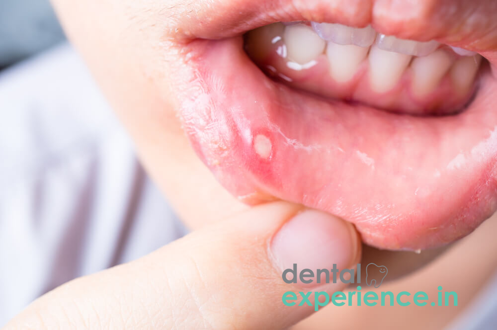
Ulcers of the lips result when the tissue layer called the epithelial covering of the labial mucosa or vermilion is broken at a single site to the point that it extends beyond the basement membrane and into the submucosa. Based on their clinical manifestation within an oral medicine context, lip ulcers can be classified as either minor traumatic injuries with a benign course, systemic autoimmune conditions (e.g., Behçet's syndrome), or even early-stage labial squamous cell carcinomas. Lip ulcers cause considerable pain due to exposure of the submucosal nerve endings that are innervated by the branches of the mental nerve (or upper labial branches of the infraorbital nerve if affecting the upper lip); therefore, they can create considerable difficulties for eating and speaking.
Clinical Definition: An open lesion in the mucous membrane that lines the lower lip which exposes the submucosal nerve endings.
Red-Flag Signs/Symptoms: A non-healing ulcer (lasting >14 days), severe unexplained pain, localized firmness (induration), or neck swelling requiring prompt medical/dental evaluation.
Emergency Indications: Facial swelling, high fever, inability to swallow liquids, or severe tissue trauma requiring immediate evaluation (within 24 hours).
Recovery Pathways: Corticosteroid topical ointments/creams, antimicrobial mouthwashes/rinses, diet modification, or laser phototherapy.
Appearance: The visual appearance is a round or oval-shaped crater in which there is a yellow-white fibrinous area within the crater. Surrounding the crater is a raised reddened area of inflammation.
Pain/Sensation: Sharp, burning sensations are experienced when consuming foods that are either warm, salty, or acidic.
Tissue Changes: Swelling of the labial tissue surrounding the lesion results in decreased mobility and thus discomfort with speech and chewing.
You should seek an immediate medical evaluation if you see:
The duration of your symptoms has lasted longer than two weeks and will continue to persist without decreasing in size.
There are changes in the physical appearance of the ulceration including induration of the skin on one side of the ulcer and/or development of pain-free lesions with rolled borders.
Presence of systemic disease signs such as unexplained fevers; weight loss; or chronic lymphadenopathy (chronic swollen neck lymph nodes).
Traumatic Injury (Thermal & Mechanical): The oral ulcers can be caused through accidental biting of the lips. Ill fitting dentures, hot food items, such as black tea, and extremely spicy food items.
Immune System Dysregulation: The immune system dysregulation that is seen with recurrent aphthous stomatitis (RAS) may be due to an abnormal response to a variety of factors including chronic stress, poor sleep, and/or minor trauma to the local tissues.
Viral Reactivation: Local viral reactivation of HSV-1 leads to painful labial vesicles known as cold sores (unlike non-viral aphthous stomatitis, commonly known as canker sores).
Chemical Irritation: Chemical irritation of the mouth lining. Some common chemical irritants are SLS found in many toothpastes, local irritation from chewing betel nuts or smoking.
Nutrient Deficiency: Nutrient deficiencies associated with low levels of vitamins B12, iron, folic acid or zinc. These nutrient deficiencies are commonly due to poorly balanced diets.
Consumption of Acidic Foods/Drinks: Consumption of large amounts of acidic local fruits (raw mangoes, tamarinds etc.) and/or very intense spiced gravies.
Systemic Diseases: Patients who have celiac disease, inflammatory bowel diseases (ulcerative colitis/Crohn's disease), or Behçet's syndrome.
Sharp Teeth/Fractured Teeth & Orthodontic Appliances: Fractured sharp teeth or orthodontic appliances rubbing on the inside of the lower lip.
Thoroughly Wash your hands with soap and water prior to handling the mouth area.
Observe in front of a lighted mirror. Place yourself in front of an adequate light source, preferably daylight, when you are going to assess a lesion.
Pull the corner of the lower lip down (or upper lip up) away from the gum line with your clean fingers.
Assess the shape of the edges of the lesion, its color and how deep it is by looking carefully at it but do not pick at the fibrin base.
The lesions caused by aphthae will feel soft and pliable to the touch. If there is a lesion that feels stiff, hard or has a lumpy base, do not delay seeking medical care immediately.
Clinical Evaluation: Visualize the margin of the lesion, its depth, and the extent of the anatomy involved through direct observation of the lesion utilizing intense illumination within the dental operative environment.
Palpation: Gently perform a manual palpation of the lesion to detect evidence of induration (stiffness) or base invasion.
Microbial/Cytologic Testing: Obtain swabs for PCR testing of viral DNA (Herpes Simplex Virus Type I) or culture tests for opportunistic infections (e.g., Candida).
Biopsy: Obtain tissue under local anesthesia if an ulcer fails to heal after fourteen days; all ulcers failing to heal may indicate the presence of oral squamous cell carcinoma or an autoimmune condition such as pemphigus vulgaris.
Mild (Minor): Less than 1 cm in diameter, shallow in the mucosa and heals without scarring in approximately 7-10 days.
Moderate (Major): Over 1 cm in diameter, at a deeper level of the submucosa; it is painful and will take approximately 2-6 weeks to heal with possible mild scarring.
Severe (Complex / Malignant Suspicion): Persists for longer than two weeks, either has no pain or forms a large, deep crater that has firm borders, is accompanied by swelling of the lymph nodes, or rapidly expands.
Anti-inflammatory Ointments: Clobetasol propionate gel (0.05%) or triamcinolone acetonide 0.1% oral paste can be applied topically two to three times daily to reduce swelling.
Mucosa-Coating Agents: Benzocaine topical anesthetics, hyaluronic acid gels, and sucralfate suspensions create a protective film over mucosal surfaces to shield sensitive tissue from irritation.
Photobiomodulation / Low-Level Laser Therapy: Direct application of low-level laser phototherapy is used in dentistry to stimulate tissue repair and provide immediate pain relief.
Systemic Therapies: For severe recurrences, oral vitamin B-complex supplementation, zinc lozenges, or a short course of systemic corticosteroids may be prescribed.
Outpatient Consultation Fees: Typically range between ₹300 and ₹700 in general clinics across metropolitan regions of Kerala (such as Thiruvananthapuram, Kochi, and Kozhikode).
Prescription Gels & Rinses: Topically applied corticosteroids and antimicrobial rinses generally cost between ₹150 and ₹500.
Low-Level Laser Therapy: Advanced photobiomodulation treatments usually cost between ₹1,000 and ₹2,500 per session.
General Coverage: Outpatient mucosal care is primarily an out-of-pocket expense. However, inpatient surgeries or major biopsy procedures may be covered under state health schemes or private medical insurance.
General Dentist/Physician: Handles primary assessment, straightforward mechanical ulcers, and small aphthous lesions.
Oral Medicine Specialist: Assesses chronic, recurrent, autoimmune, or unusual oral and mucosal disorders.
Dental/oral/maxillofacial surgeon: Conducts biopsy evaluations, removal of potentially malignant areas, reconstructive surgery for major, extensive destruction to deep tissues.
Emergency (Within 24 hours): Swelling in the face, extremely high temperature, unable to swallow liquids due to the ulceration and a significant traumatic disruption of the skin/tissue layers.
Urgent (within 2-3 days): Very painful ulcers causing malnutrition/nutrient deficiencies or an acute outbreak of a virus with a severe onset.
Routine/Elective (Scheduled within 1-2 weeks): Regular follow-up on very slow healing ulcers and recurrent, mild aphthous lesions.
Why am I getting recurrent lip ulcers after consuming spicy Kerala food?
While the spicy components in your curry (chili/tamirand) are irritant chemicals that can exacerbate pre-existing trauma to the lip mucosa and stimulate an inflammatory response; they do not directly induce ulcers.
Will my lip ulcer develop into mouth cancer?
Traumatic or common aphthous ulcers will not transform into cancer. However, early stage labial squamous cell carcinoma may mimic benign appearing ulcers. Therefore, any ulcer longer than two weeks should have consultation from a medical professional.
What at-home treatments provide relief for sore inside of the lower lip?
Warm saline rinse (or diluted coconut oil - "Oil Pulling") provides temporary soothing to the damaged tissues. Do NOT apply raw spice or other harsh antiseptic compounds to the open wound - these will burn your skin and create more damage.
Chronic lip ulcers are commonly viewed as small annoyances. However, the continued presence of ulcers may indicate systemic distress and/or localized pathology. Prolonged friction to the mucosa due to damage to teeth, the use of harsh oral care products, or a lack of dietary control will extend the time required for healing. Early evaluation by a medical or dental professional for all lesions that have existed beyond 14 days is important to quickly treat the pain associated with these ulcers, reduce potential scarring of the involved tissue, and assess the extent of possible mucosal disease through appropriate diagnostic testing.














.jpg)
.jpg)

.jpg)
.jpg)

.jpg)
.jpg)
.jpg)
.jpg)
.jpg)
.jpg)
.jpg)
.jpg)