Asymmetries in the gum-line refer to the area where the gingiva (the tissue around your teeth) is irregularly shaped as it surrounds a tooth. This means some areas have less gum than they should for aesthetic purposes and other areas have too much gum for both aesthetically pleasing results and proper periodontal health. Gingival recessions can compromise both dental aesthetics and periodontal health by exposing a portion of the root surface of the tooth, providing an environment where harmful bacteria can reside. Additionally, excessive gum will create a deeper pocket, which will also serve as a reservoir for pathogens.
Clinical Definition: Gum line that does not appear symmetrical due to growth of soft tissue at one location compared to another location, focal loss of attachment of the gingiva, abnormal shape of alveolar bone, or the tooth itself.
Indications For Urgent Treatment: Bleeding from the mouth without trauma to the area, pain or tenderness to touch, spontaneous bleeding from a tooth with no apparent reason, mobility of the teeth, or separation of gum from tooth.
Treatment Options: Gingivoplasty via laser, Crown Lengthening (Crown Exposure), Pinhole Surgical Technique, Orthodontic Alignment.
Timeframe Of Recovery: The body heals most rapidly in 7–14 days; Complete bone remodeling may take 3–6 months.
The symptoms most commonly associated with patients who have an abnormal height differential of the gums are both visually observable and physically felt by the patient throughout their smile:
Visual Height Differential: Some teeth will be significantly shorter or longer than the adjacent teeth when a patient is smiling.
Visible Exposure Of Root Structure:Yellowish coloration of cementum exposed at the tooth base as a result of localized recession.
Localized Swelling And/or Redness: The inflamed gingiva is usually dark pink to red in appearance as it surrounds certain teeth.
Tenderness When Brushing Or Flossing: Patients may feel tenderness when they brush and/or floss their gums over areas where the unevenly formed gingiva exists.
Food Impaction: Many times food such as cassava or red meat becomes lodged into the deeper crevices surrounding the unevenly formed gingiva.
You should seek emergency periodontic care immediately if you experience:
A constant, unrelenting flow of pus from the gumline when you apply gentle pressure.
Increased mobility of one or multiple teeth, or any change in your occlusal relationship.
Exposure of more tooth surface area that has become sensitive, accompanied by persistent, sharp pain.
Any type of bleeding which continues despite lack of mechanical trauma to the gums.
The unevenness in gum line is caused by a combination of structural, mechanical, and biological issues that affect how evenly the gum lines grow back as your teeth mature:
Altered Passive Eruption: The passive eruption process is where the gum recedes away from the roots of the teeth as they mature. If this does not occur it will leave excessive amounts of gum overlying the enamel matrix (the outer protective layer of the tooth).
Localized Periodontal Disease: When bacterial plaque builds up and causes inflammation to break down the alveolar bone supporting the teeth, it creates an uneven surface for the gum to sit upon.
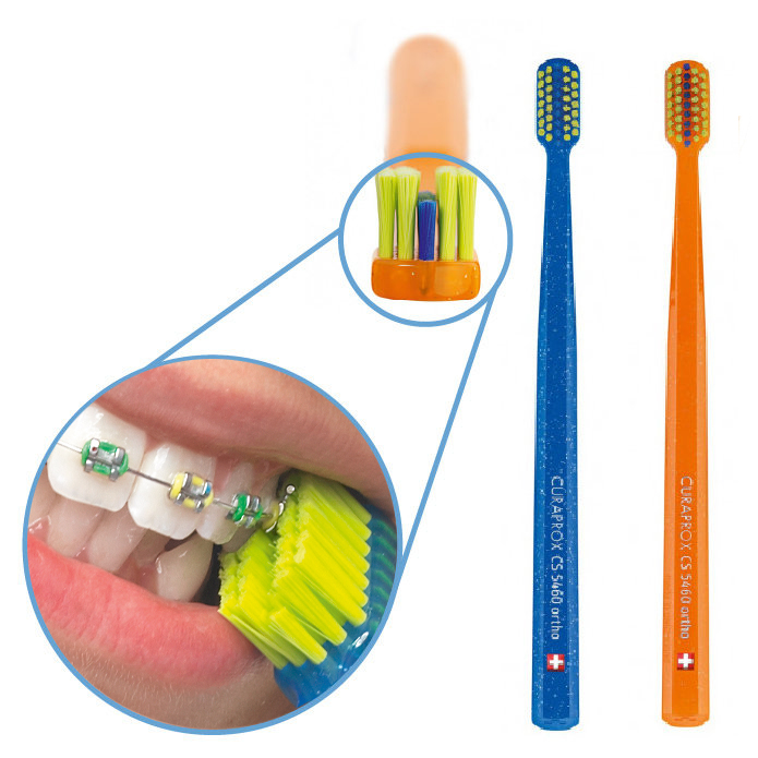
Aggressive Oral Hygiene Habits: Brushing with hard bristles or scrubbing too aggressively with cross brush strips can be detrimental to the health and integrity of the delicate gum margins.
Malocclusions and Dental Trauma: Teeth that are malpositioned relative to each other apply uneven pressure when you chew. This can accelerate recession at specific sites along the gum margin.
Para-Functional Habits: Grinding your teeth severely at night (bruxism), puts flexural stress on the tooth neck which can create abfractions (a lesion where the tooth has been worn away and the enamel has fractured off) and cause recession in the gum margin.
There are several systemic risk factors and lifestyle choices that make people more vulnerable to developing uneven gum contour:
Diet: Consuming foods that are difficult to chew such as dried fish or raw jaggery frequently enough, can cause microtrauma to the soft tissue margins of your mouth.
Genetic Predisposition: Thin biotype gingiva makes you more susceptible to recession due to slight mechanical irritation.
Hormonal Fluctuations: Menopause, pregnancy, etc., create an environment of increased inflammation response to low-grade plaque.
Tobacco Use: Smoking or using betel nuts damage the small blood vessels in the soft tissues, creating conditions for rapid loss of the soft tissue at irregular intervals.
Visual assessments can be performed by yourself in your home environment using an unbroken mirror, along with sufficient light.
Clean Your Hands Before Touching Your Lips or Oral Cavity: Clean your hands with a liquid soap, and then touch the area of your lips or oral cavity you wish to examine.
Lift Upper Lip & Lower Lip: Carefully draw up on the top of your mouth using both thumbs and fingers. Then gently pull back on the bottom of your mouth while keeping your teeth slightly apart.
Examine the Shape of the Gum Line: Look closely at the highest part (the "peak") of the gum line directly above each of your teeth. The healthy gum lines typically exhibit a somewhat symmetrical, curved shape.
Find the Discrepancies: Look at each tooth to find if the gum line is located below or above its neighboring tooth(s). If it's significantly lower or higher, there may be some type of issue.
Gently Press Against Tissue Near Tooth Base: Use your index finger and press against the tissue surrounding the tooth base to check for swelling. You should also look for redness and/or tenderness.
Warning: Never use metal instruments, toothpicks or other sharp objects to probe the gum space. Doing so could cut through the periodontal ligaments.
A periodontist will utilize precise diagnostic methods to create detailed images of soft tissue contour and underlying bone architecture:
Periodontal Probing: The periodontist utilizes a standardized measuring device called a periodontal probe to measure how far the gum tissue is away from each tooth. All measurements are taken in millimeters; anything above 3 mm indicates either inflammation or bone loss.
Digital Radiographs (Intraoral X-Ray): Each tooth has a unique structure consisting of a tooth crown, neck, and root. This structure allows digital radiographs (x-rays) to provide a view into the alveolar bone height surrounding each root. Digital radiographs help determine whether you have a problem associated solely with the tissue or one related to structural bone problems.
Cone Beam Computed Tomography (CBCT): This advanced 3D technology provides a non-invasive way to evaluate buccal bone thickness before performing any surgical procedures such as recontouring.
Vitality Testing: Thermal tests (hot/cold test) and electrical tests (electrical pulp test) assess the health status of each tooth by verifying that all visible exposed areas of teeth do not represent internal necrotic pulp chambers pushing them out.
|
Severity Level |
Clinical Findings |
Primary Cause |
Typical Treatment Strategy |
|
Mild (Stage I) |
1–2 mm zenith height variation; intact interdental papillae |
Minor altered passive eruption or localized friction |
Soft tissue laser gingivectomy |
|
Moderate (Stage II) |
3–4 mm asymmetry; localized root exposure or 4–5 mm pockets |
Partial bone height variation, localized recession |
Micro-surgical recontouring & scaling |
|
Severe (Stage III) |
>4 mm height difference; severe bone loss or root exposure |
Advanced periodontitis, severe malocclusion |
Flap surgery, bone grafting, or orthodontics |
Modern periodontology as it relates to minimally invasive treatments to create a symmetrical appearance includes:
Diode / Er:YAG Laser Gingivectomy: The Diode/Er:YAG laser allows for precise gingival excision by using high energy pulses that vaporize the unwanted tissue along with the elimination of harmful microorganisms; and simultaneously seal the capillary bed at the same time eliminating bleeding and requiring no sutures during the post operative process.
Esthetic Crown Lengthening: This combines the reduction of soft tissue with micro-contouring of the underlying bone creating a permanent lowering of the tissue levels over short teeth.
Pinhole Surgical Technique (PST): Using no scalpels, this procedure uses tiny entry points to loosen the tissue and slide it down over exposed roots, supported with collagen membrane.
Connective Tissue Grafting: Connective tissue grafts are placed via micro-surgery from the palatal donor sites into areas deficient of lost gum height around severely receding teeth.
The cost of treatment in Kerala varies depending upon the complexity of surgery, whether laser was used, type of materials selected:
|
Treatment / Procedure |
Cost Range (INR / ₹) |
|
Laser Gingivectomy (per quadrant) |
₹3,500 – ₹8,000 |
|
Esthetic Crown Lengthening (per tooth) |
₹4,000 – ₹9,500 |
|
Pinhole Surgical / Grafting Technique (per site) |
₹12,000 – ₹25,000 |
|
Multi-tooth Comprehensive Recontouring |
₹25,000 – ₹60,000 |
Note: Purely cosmetic gum leveling will typically have to be paid personally. Insurance companies covering comprehensive private medical plans or corporation dental benefit programs covering structure of bone loss or serious periodontal pocketing may cover costs of these procedures throughout the state of Kerala.
Selecting the right dental specialists can help ensure your treatment will produce both aesthetically pleasing and functionally successful results:
General Dentist: Can diagnose an area of asymmetry; perform scaling and periodontal debridement; use a soft-tissue CO2 laser for minor tissue sculpturing.
Periodontist: Should be selected as the first specialist if there are areas that need to have advanced tissue grafts performed; if you need bone recontoured to create symmetry; or if you require pinhole surgery.
Prosthodontist: Prosthodontists should be selected in conjunction with a general dentist, especially when creating uneven gum levels along with either a full mouth rehabilitation or veneers/crowns.
Orthodontist: Orthodontists are recommended when you need to move teeth (extrude/intrude) to make up for differences in gum level heights that cannot be corrected by surgical means.
The timeline for treatment depends upon the reason(s) why you have an asymmetric condition:
Emergency (within 24–48 hours): Severe pain; active swelling; or pus draining from the site of a very high gum margin which requires immediate abscess drainage.
Rush (2–4 weeks): Rapidly developing/progressing exposed roots or large pockets resulting in significant temperature sensitivities.
Routine / Elective (at your convenience): Asymmetric soft tissue contouring without active bone loss; inflammation; or pain.
Why do my gums remain uneven after a daily routine of brushing your teeth?
The regularity of brushing will never correct for a pre-existing condition due to differences in the thickness of the gingiva (genetic), position of the teeth (anatomical) or from using an overly vigorous approach to cleaning the teeth (mechanical). Brushing aggressively can cause uneven tissue loss on both sides.
How painful is it to have laser gum contouring done? And how long before you recover?
You won't experience any discomfort during laser contouring as we numb the area first. Some mild soreness can last anywhere from 48 hours to four days. Soft tissue should be fully healed in about ten to fourteen days.
Will uneven gums grow back after they have been surgically removed?
Any soft tissue that has been reshaped to be level with the bone will not grow back. If there were areas of bone where the soft tissue had receded but then additional soft tissue was removed to get rid of excess, the soft tissue will continue to shrink over time.
To correct uneven gum levels we must evaluate both soft tissue and bone structure. Today there are many minimally invasive treatments that can help create a symmetrical look by contouring the gums with a diode laser or through a procedure called "pin hole" surgery. To maintain long-lasting results in function and aesthetics you must first treat all underlying inflammation before doing any aesthetic gum changes.


.jpg)
.jpg)
.jpg)
.jpg)
.jpg)
.jpg)
.jpg)
.jpg)
.jpg)
.jpg)
.jpg)
.jpg)
This site is part of the DEC Dental Network — a Dental Equity & Connectivity ecosystem of clinical, educational, and patient-support brands operated by Team Dental Pvt. Ltd. Our network works as one — guiding you from your first search to your final smile.
© 2026 Team Dental Pvt. Ltd. · Founded 2013, Angamaly, Kochi, Kerala
About DEC Dental