.jpg)
White spot lesions are small areas where the enamel is broken down by acid from food and drink or as a result of the oral biofilm. They appear clinically as white, chalky patches on the tooth. These are examples of non-carious defects in enamel or early caries in enamel (ICDAS Code 1 & 2). White spots occur due to the leaching of calcium and phosphorus from the hydroxyapatite crystals that make up the structure of enamel by acidic substances produced by dental plaque bacteria and/or continuous chemical exposure. If left untreated, they will cause structural weakening of the enamel resulting in potential sensitivity and decay to the dentin and pulpal chamber underneath.
Clinical Definition: Loss of mineral content within the enamel with resultant reduction in density and white discoloration visible upon inspection.
Indicators of Urgency: The rapid increase in size of the chalky areas; the roughness of the area; localized sharp pain at specific times; or micro pitting of brown color.
Treatment Options: First-line treatments for white spot lesions include microabrasion; resin infiltration; application of CPP-ACP for topical remineralization; and porcelain veneer treatment.
Visual Changes: Enamel appears chalky, milky, white with bands or cloudy patches on the smooth labial surfaces of upper and lower incisors.
Texture Differences: Surface texture transitions from a very smooth, glassy enamel to a slightly rougher, matte, or porous surface structure.
Sensitivity to Temperature: Sharp pains due to exposure to cold temperatures (drinks) or hot temperatures (hot tea).
Changes in Color: White spots become stained and darker brown in color over time.
Prompt clinical action is required for structural damage to the enamel including microfractures, crumbling tooth edges, pinpoint cavities, etc. Porous areas causing constant pain while chewing indicate irritation at deeper levels of the tooth close to the pulp chamber. If there are white spots and bleeding gums or localized darkening of the gums adjacent to a white spot, an immediate examination should be performed to assess possible rampant cervical decay or active periodontitis.
Fluorosis of Teeth: The ingestion of excessive amounts of fluoride by an individual during their developing teeth stage (from birth to age eight). This can disrupt the function of the ameloblasts (the cells that form the enamel of a tooth) and lead to an alteration in the structure of the enamel matrix crystalline structures.
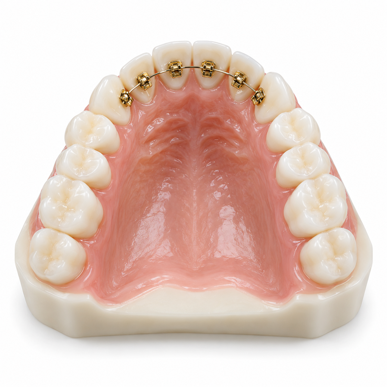
Decalcification: The inadequate removal of plaque from the margin areas surrounding the brackets used for fixed appliance treatment. This allows the local area to accumulate and be damaged by acidic substances.
Hypoplasia of Enamel: The insufficient production of the enamel matrix at some point during the process of tooth development due to either a high fever while a child is still developing, poor nutrition, or other systemic disease processes occurring during this time frame.
Eroding Effects of Acidic Diet: The frequent consumption of foods and beverages which are highly acidic such as tamarind (puli), citrus-based curry preparations, carbonated soft drinks, and fermentable carbohydrate products.
Sources of Water / Geographic Areas: Deep bore wells with untreated water in high-fluoride geographic locations throughout Kerala (parts of Palakkad, Alappuzha, Kollam) are being utilized as the source of drinking water.
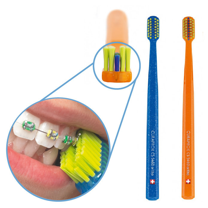
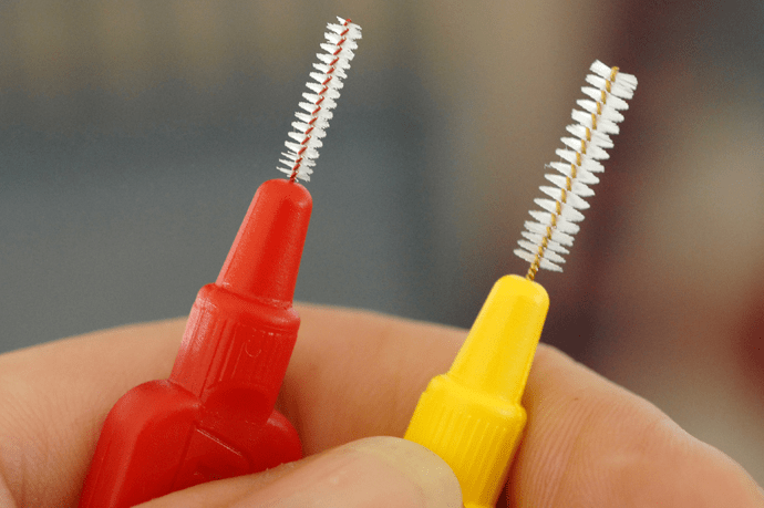
Failure to Maintain Good Oral Health Practices: A lack of consistent good oral hygiene practices resulting in failure to regularly clean away the sticky biofilm which forms around orthodontic appliances or in the cervical region of the teeth.
Reduced Saliva Flow / Xerostomia: As a result of medication use or systemic medical conditions; saliva provides protection against acids and helps provide natural remineralizing ions for dental tissues.
Young Children’s Habitual Swallowing Behavior: Repeatedly swallowing amounts of fluoridated toothpaste in young children during early stages of dentition development.
Perform a preliminary self-check:
Clean and Dry: Gently brush your front teeth using a soft-bristled toothbrush to clean all surfaces. Use a soft tissue to wipe the front surface of each tooth so it is completely free of moisture.
Lighting and Mirror: Position yourself in the direct path of a strong source of light (usually overhead), facing a mirror so you can see the entire dental arch.
Visual Examination: Inspect if the white areas appear as continuous, cloudy bands on the front surfaces of your front teeth (typical of fluorosis) or as isolated, small areas toward the gumline (possible early stages of decay).
Important Safety Warning: Avoid the use of any metal instruments such as metal picks, toothpicks, or other sharp objects to investigate the area since they may cause damage by crushing weakened, demineralized enamel.
The process used by dentists to diagnose includes visual examination and tactile assessment techniques based upon guidelines established by ICDAS criteria, along with gentle drying of the surface of the enamel:
Transillumination: The use of very high-intensity light produced from a fiber-optic lamp, which shines through the tooth structure to determine the thickness of the lesions and differentiates structural defects from superficial staining.
Radiographic Diagnostic Technique: Digital radiographs in the form of bitewing X-rays will confirm whether or not the demineralization penetrated to the enamel-dentin junction (EDJ).
Advanced Imaging Technology: QLF (Quantitative Light-Induced Fluorescence) diagnostic technology was developed specifically to evaluate the extent of mineral loss from the tooth structure, and therefore monitor changes in remineralization.
Mild (Superficial): White specks or faint white lines on the surface of teeth which cover <25% of the total surface area. Surface enamel remains intact and smooth.
Moderate (Sub-Surface Loss): Distinct chalky white patches which cover up to 50% of the tooth surface. Minor surface scratches are present but no cavitation occurs.
Severe (Structural Compromise): Multiple white patches, along with pitting of the enamel, crumbled surfaces, secondary browning, and localized hypersensitivity occur throughout the affected tooth.
Topical Remineralization: Use of CPP-ACP / Tooth Mousse or high-concentration fluoride gels to repair the mineral lattice structure within superficial lesions.
Enamel Microabrasion: A combination of mechanical abrasion and mild acid application to remove opacity from shallow sub-surface areas (up to 100 microns deep).
Resin Infiltration (Icon Protocols): The Icon method utilizes a light-cured low-viscosity resin to penetrate into porous enamel micro-cavities that are produced by white spot lesions. By matching the refractive index of enamel, this method allows for natural masking of white spots.
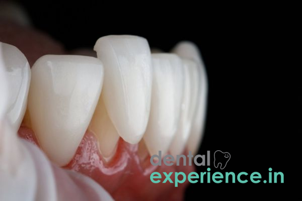
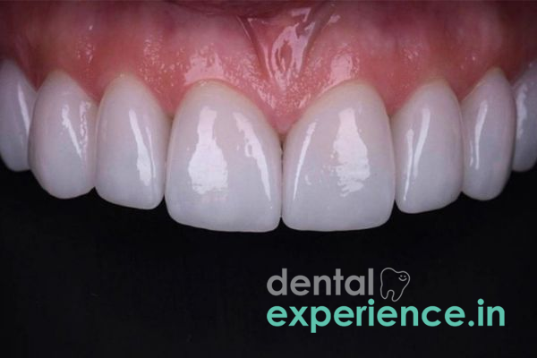
Direct Composite Bonding / Porcelain Veneers: Directly bonding lab-fabricated porcelain veneer shells or layering composite material directly over severely damaged teeth when enamel thickness has been compromised.
Diagnostic testing and treatment costs in Kerala will be based on the average cost of dental services in this region of India:
Fluoride Varnish and/or Remineralization Therapy: ₹800 – ₹2,000 per visit.
Treatment Using Microabrasion: ₹1,500 – ₹3,500 per tooth.
Icon Resin Infiltration (Icon Therapy) Treatment: ₹4,000 – ₹9,000 per tooth.
Aesthetic Ceramics (Veneers): ₹10,000 – ₹22,000 per tooth based upon the quality of ceramic used.
General Dentists: General dentists can perform clinical examinations, radiographic examinations (diagnostic imaging), topical fluoride applications, and microabrasion treatments.
Pediatric Dentists / Pedodontists: Pediatric dentists/pedodontists have specialized training and expertise that focuses on treating children who experience developmental fluorosis, enamel hypoplasia, and early childhood caries.
Cosmetic Dentists / Prosthodontists: Cosmetic dentists/prosthodontists have extensive experience managing complicated multiple-tooth aesthetic case scenarios which require either advanced Icon Resin Infiltration, comprehensive bonding techniques, or high-end aesthetics from ceramic veneers.
Emergency Appointment (Within 24 – 48 Hours): Sharp pain along with new white spot development; surface crumbling; brownish micro-fracture visible.
Scheduled Appointment (2 – 4 Weeks): Scheduling an appointment for follow-up consultation 2 to 4 weeks after braces or when cosmetic improvement is desired on existing fluorosis white spots.
Why do we see white spots appear in some areas of the state of Kerala?
The high levels of naturally present fluoride in the water table throughout much of both the inland and coastal regions of Kerala contribute to the formation of developmental dental fluorosis in young children. This results in the appearance of whitened opaque patches in their enamel. These patches can resemble "chalk" or "paint".
Are white spots which occur as a result of environmental exposure to fluoride able to be removed through brushing with regular toothpaste?
No. White spots are structural defects caused by deficiencies of minerals that make up the enamel matrix, rather than stains on the surface of your teeth. Using hard-bristled brushes or abrasive toothpastes may further weaken already fragile enamel. Additionally, this will likely increase discoloration due to abrasion.
How long does resin infiltration treatment take, and is it painful?
Resin infiltration typically takes 45 to 60 minutes in a single appointment. The procedure is entirely non-invasive, requiring no drilling or local anesthesia.
White spots on the tooth surface require an organized diagnostic examination to determine whether or not to perform restorative treatment. Non-invasive methods for conserving minerals are preferred (e.g., microabrasion combined with resin infiltration) prior to performing an invasive restoration. In addition to using controlled fluoridated drinking water sources and maintaining a balanced diet, patients will experience long-term stable aesthetics as well as healthy enamel structure in fluoride-rich areas.




.jpg)
.jpg)
.jpg)
.jpg)
.jpg)
.jpg)
.jpg)
.jpg)
.jpg)
.jpg)
.jpg)
.jpg)
This site is part of the DEC Dental Network — a Dental Equity & Connectivity ecosystem of clinical, educational, and patient-support brands operated by Team Dental Pvt. Ltd. Our network works as one — guiding you from your first search to your final smile.
© 2026 Team Dental Pvt. Ltd. · Founded 2013, Angamaly, Kochi, Kerala
About DEC Dental