
A "dark" tooth after a facial injury or blow to the face represents the destruction of the internal structures that support the teeth. After a tooth is subjected to mechanical stress, it can cause rupture of blood vessels inside of the pulp chamber, which houses all of the nerve tissue and vascular supply. The breakdown of hemoglobin into iron compounds leaches into dentin via dentinal tubules located below the enamel matrix; this causes an intrinsic color change on the surface of the tooth to be gray, brown, or black. A post-trauma dark tooth is categorized with other forms of tooth discoloration under the umbrella of intrinsic chromatic changes due to pulpal necrosis, internal resorption, or pulpal obliteration.
Clinical Definition: Tooth discoloration due to internal hemorrhage, nerve death, or pulpal obliteration following a traumatic injury to the tooth.
Indications for Emergency Treatment: Sudden severe pain without provocation, localized swelling at the gum line, pus drainage from the gum line, or sudden looseness of the tooth.
Treatment Alternatives: Internal bleaching of non-vital teeth, root canal / endodontic therapy, porcelain laminate veneer(s), full-coverage ceramic crown(s).
Signs of Post-Trauma Dark Tooth Syndrome include the following visible & tactile symptoms that may evolve over several months after the traumatic event:
Visual Signs: A tooth normally a light off-white or yellowish color will darken through weeks or months as it progresses through a series of colors ranging from slate grey to blackish-brown.
Physical / Tactile Signs: The tooth will be tender to touch with a blunt object, tender when chewing or biting down forcefully on hard foods, or lose normal temperature sensitivity due to nerve tissue degeneration.
Gum Tissue Changes: Redness of the gums surrounding the traumatized tooth, swollen tissue for an extended period of time, and/or a fistula (a small gumboil or sinus tract at the base of the tooth).
Any of the following severe signs indicate an immediate need for clinical intervention:
Acute Throbbing Pain: Pain radiating to the jaw, ear, or neck, indicating an infection spreading through the pulpal canal.
Active Suppuration: Open sores or pus discharge along the gumline accompanied by a persistent foul taste or odor (halitosis).
Facial Asymmetry: Asymmetrical swelling of the face, lip, cheek, or lower jaw region.
Tooth Mobility: A loose feeling or displacement of the traumatized tooth in its bony socket.
Post-traumatic tooth darkening is caused by specific mechanical and biological disruptions within the internal pulp chamber:
Direct Mechanical Force: A direct blow to the face or mouth (such as during contact sports, falls, or accidents) severs or ruptures the delicate neurovascular bundle at the root apex, leading to pulpal hemorrhage and tissue necrosis.
Masticatory Trauma (Severe Biting Stress): Biting down forcefully at an unfavorable angle on hard substances (such as uncracked nuts or hard seafood shells) can cause micro-fractures in the dentin or crush internal pulpal tissues, interrupting normal blood flow.
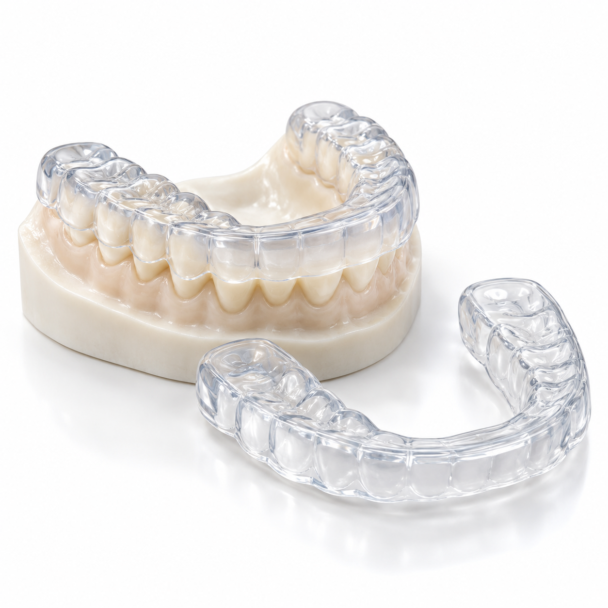
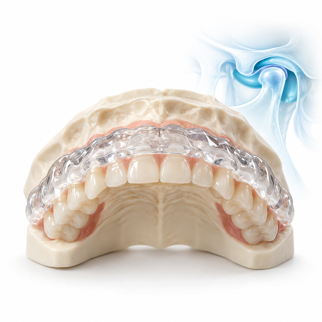
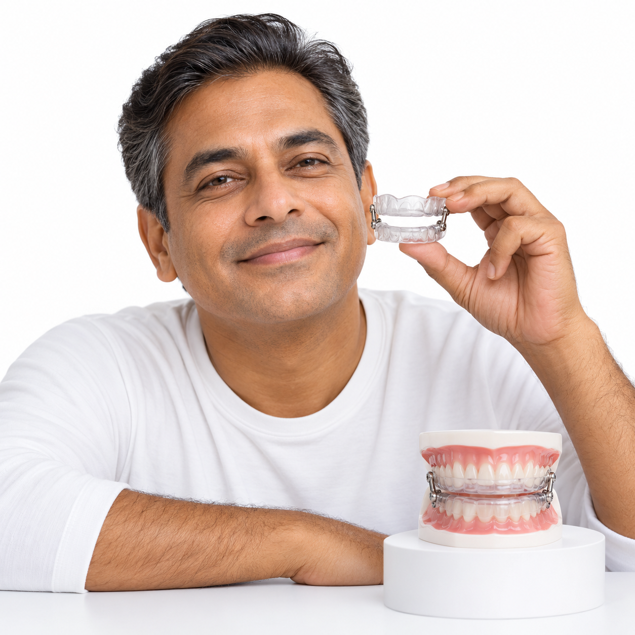
Micro-Trauma (Parafunctional Habits): Repetitive strain from chronic bruxism (teeth grinding) or jaw clenching exerts continuous excessive stress on the periodontal ligament and pulpal blood vessels, potentially triggering calcific metamorphosis or pulpal death over time.
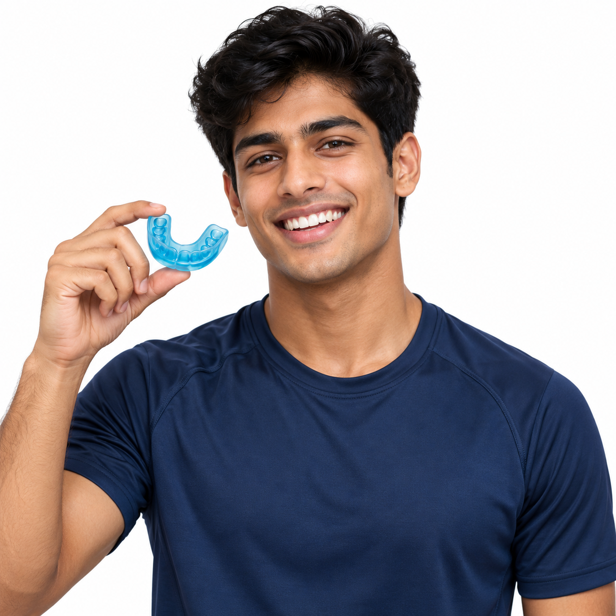
Participation in Contact Sports: High-impact athletics without custom mouthguards significantly increase the likelihood of dental trauma.
Pre-Existing Structural Weaknesses: Teeth with large composite restorations, deep dentinal caries, or enamel dysplasias are more susceptible to pulpal disruption under impact.
Younger Age (Children & Adolescents): Children and young adults have significantly larger pulp chambers with wider apical foramina, making their teeth more susceptible to internal vascular rupture following trauma.
Perform this safe, non-invasive home check in front of a well-lit mirror:
Visual Examination: Compare the color of the discolored tooth with that of adjacent teeth while viewing both in good light. Identify any differences in color.
Light Transmission Test: Shine a small penlight through the lingual surface of the tooth. Teeth which are non-vital or necrotic will appear opaque and dark, whereas healthy teeth allow even light transmission.
Examination of Soft Tissues: Lift your upper lip and examine the soft tissues overlying the roots of the teeth. Look for red areas, swelling, or small blisters/fistulae above the root tip.
Evaluation of Mobility: Gently place light finger pressure against the crown of the tooth. Do not pull or wobble the tooth forcefully. The tooth should feel solidly fixed into its alveolar socket.
A dentist utilizes standardized clinical diagnostics in conjunction with one another to assess pulpal vitality and structural integrity:
Digital Radiography: Intraoral periapical (IOPA) radiographs identify apical periodontitis, root fractures, alveolar bone loss, and internal or external root resorption.
Electric Pulp Testing (EPT): Measures the response threshold of internal nerve fibers when low-level electrical currents are applied.
Thermal Vitality Testing: Applies cold stimulus (endodontic ice spray) to assess pulpal nerve responsiveness and vitality.
Periodontal Probing: Evaluates localized deep pocketing to rule out vertical root fractures.
|
Grade Level |
Structural State |
Clinical Characteristics |
Primary Intervention |
|
Grade 1 |
Superficial Discoloration |
Intact pulp, transient hyperemic staining. |
Observation, external vital monitoring. |
|
Grade 2 |
Pulpal Obliteration |
Calcific metamorphosis; canal narrows and the tooth turns yellow or amber. |
Long-term clinical monitoring or external cosmetic restoration. |
|
Grade 3 |
Pulpal Necrosis |
Complete nerve death; gray/black discoloration; asymptomatic or mild ache. |
Root Canal Therapy + Internal Walking Bleach / Veneer. |
|
Grade 4 |
Periapical Infection |
Infection spreads to bone; periapical abscess, severe swelling, high tooth mobility. |
Urgent endodontic debridement, infection drainage, or surgical extraction if non-restorable. |
Modern dental restorations emphasize preserving pulp function, restoring structural integrity, and achieving optimal aesthetics:
Internal Bleaching (Walking Bleach): Applies a sodium perborate or hydrogen peroxide gel inside the pulp chamber of an endodontically treated non-vital tooth for several days to oxidize internal iron compounds and blood breakdown products within the dentinal tubules.
Root Canal Treatment (RCT): Cleanses, disinfects, and obturates a necrotic canal space using bioceramic sealers and gutta-percha, preventing bacterial colonization of the root system.
Porcelain Veneers: Thin ceramic shells bonded to the front surface enamel to mask persistent dark staining while preserving maximal natural tooth structure.
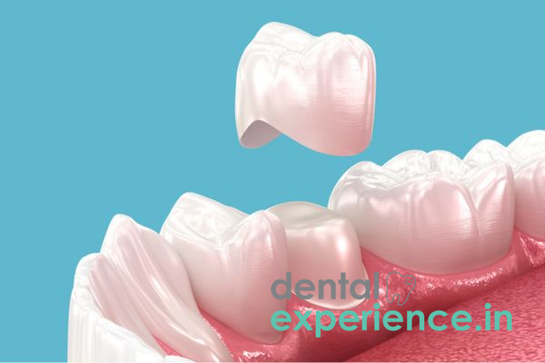
Zirconia or E-Max Crowns: Full-coverage ceramic crowns utilized when the tooth's structural integrity has been severely compromised by trauma or deep restorations.
Treatment pricing in Kerala varies based on clinical complexity, chosen materials, and practitioner expertise:
|
Procedure |
Estimated Cost Range (INR / ₹) |
Primary Cost Drivers |
|
Root Canal Treatment (Front Tooth) |
₹3,500 – ₹8,000 |
Rotary instrumentation, bioceramic sealers. |
|
Internal Bleaching (Per Session) |
₹2,000 – ₹4,500 |
Chemical concentration, number of visits. |
|
E-Max / Zirconia Ceramic Crown |
₹8,000 – ₹18,000 |
Digital CAD/CAM milling, lab quality. |
|
Porcelain Veneer |
₹9,000 – ₹20,000 |
Custom shading, aesthetic lab technician fees. |
Note: Out-of-pocket expenses can often be offset by private dental insurance policies or regional healthcare schemes depending on hospital empanelment.
General Dental Practitioner: Ideal for initial screening, preliminary digital X-rays, emergency stabilization, and simple composite restorations.
Endodontist: Root canal specialist required to navigate calcified or narrow canals, manage pulpal necrosis, and perform internal bleaching protocols.
Prosthodontist: Reconstructive specialist trained in high-aesthetic porcelain veneers, full CAD/CAM ceramic crowns, and complex smile rehabilitation.
Urgent / Emergency (Within 2 – 4 Hours): Required when a tooth is avulsed (knocked out), severely luxated/displaced, or accompanied by uncontrolled hemorrhaging or severe facial trauma.
High Priority / Urgent (Within 24 – 48 Hours): Warranted when experiencing acute throbbing pain, soft tissue swelling, or visible pus drainage from the gumline.
Elective / Scheduled (Within 2 – 4 Weeks): Best suited for gradual, painless tooth discoloration without acute swelling or active infection.
Does a darkened tooth have a chance to become white by itself?
Only rarely. If a tooth sustains mild trauma causing transient pulpal hyperemia, temporary discoloration may occasionally clear up on its own. However, once pulpal necrosis sets in, the dark gray or black discoloration remains permanently unless professionally treated.
Do all teeth with a grayish / black color require a root canal?
No. Sometimes a discolored tooth responds normally to vitality testing (cold and EPT), indicating the nerve is still vital (such as in calcific metamorphosis). In these instances, external whitening, microabrasion, or veneers can be applied without performing a root canal.
What should I expect to pay for whitening a discolored front upper tooth in Kerala?
Total costs depend on whether the tooth needs internal bleaching, endodontic treatment, or a ceramic crown/veneer. Overall treatment plans range from approximately ₹3,500 to ₹20,000 per tooth across contemporary dental clinics in Kerala.
Early assessment of post-traumatic tooth color changes is critical to avoiding purely cosmetic treatment attempts. Post-trauma discoloration usually stems from a necrotic pulp that may be silently causing resorption of the supporting alveolar bone without causing immediate pain. Over-the-counter retail whitening products cannot treat an internally necrotic pulp and may cause soft-tissue irritation. Restoring long-term masticatory function and smile aesthetics is highly predictable with timely endodontic therapy and modern ceramic restorations produced through CAD/CAM technology.


.jpg)
.jpg)
.jpg)
.jpg)
.jpg)
.jpg)
.jpg)
.jpg)
.jpg)
.jpg)
.jpg)
.jpg)
This site is part of the DEC Dental Network — a Dental Equity & Connectivity ecosystem of clinical, educational, and patient-support brands operated by Team Dental Pvt. Ltd. Our network works as one — guiding you from your first search to your final smile.
© 2026 Team Dental Pvt. Ltd. · Founded 2013, Angamaly, Kochi, Kerala
About DEC Dental