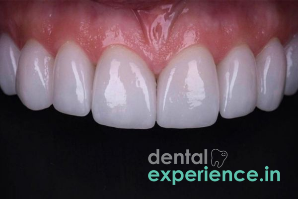
.jpg)
Dental restorations like composites, amalgams, and cements can develop discoloration over time. Discoloration is caused by superficial staining, marginal microleakage at the tooth-restoration interface, and/or the chemical degradation of the resin matrix within the restoration over time.
In terms of general dental classification, discoloration of a dental restoration is typically categorized as marginal breakdown or aesthetic degradation of a restoration. The primary cause of both marginal breakdown and subsequent discoloration is microfracturing at the tooth-restoration interface, which allows various substances from the oral environment to enter the micro-gap between the tooth and restoration. These include oral fluids, bacteria, and chromogens (coloring agents found within foods and beverages). In addition to creating an unaesthetic appearance, these substances can create a retentive site where secondary caries may develop beneath the restoration, compromising the structural integrity of the tooth's enamel and dentin matrices.
Clinical Description: Deterioration affecting the color and integrity of direct or indirect filling materials due to surface staining, marginal microleakage, or internal material degradation.
Emergency Signs/Indications: Darkened area beneath the restoration, sharp pain upon biting down, sudden temperature sensitivity, or unpleasant taste/odor near the restoration site.
Treatment Options: Surface polishing, resin sealant application, composite replacement, indirect porcelain veneers, or full-coverage ceramic/zirconia crowns.
Visual Changes: Gradual yellowing, browning, or darkening at the margins (edges) of the filling.
Tactile Changes: A rough or catchy sensation when passing the tongue over the surface of the restoration.
Marginal Staining: Darker lines or outlines defining the margin where natural enamel meets the composite resin.
Temperature & Chemical Sensitivity: Mild, localized pain in response to the consumption of sweet, hot, or cold foods and beverages.
Subsurface Discoloration: Gray or black translucency and shadow beneath the natural enamel matrix, indicating active secondary decay.
Superficial discoloration is primarily a cosmetic concern; however, there are several clinical red flags indicating deep structural compromise or ongoing bacterial invasion into the tooth structure:
Spontaneous Throbbing Pain: Spontaneous or throbbing pain that may be indicative of irreversible pulpitis requiring endodontic evaluation.
Bad Taste or Halitosis: Unpleasant taste or persistent bad breath caused by food impaction and bacterial colonization beneath a compromised restoration margin.
Tooth Mobility or Fracture: A tooth that feels loose at the gumline, exhibits abnormal mobility, or has partially broken off while chewing.
Gingival Abscess or Swelling: A small pimple-like swelling (fistula/parulis) on the gums adjacent to the discolored restoration, indicating a periapical or root canal infection.
Marginal Microleakage: Microleakage occurs when tiny spaces between the tooth and filling develop over time, allowing oral fluids and bacteria to penetrate beneath the restoration.
Chromogenic Ingestion: Consuming large amounts of dark-colored foods and beverages daily leads to surface absorption and staining of composite resin.
Material Degradation and Corrosion: Restoration materials change color over time; composite resins undergo chemical degradation of the resin matrix, while dental amalgams experience corrosion and surface tarnish, causing them to turn grey, brown, or black.
Secondary Caries: Recurrent bacterial decay forms along the margins or beneath an existing restoration.
Dental Trauma: Physical force impacting the restored tooth creates micro-fractures within the restoration or causes pulpal hemorrhage within the pulp chamber.
Dietary Habits: Regular consumption of dark beverages, such as strong tea or black coffee, and dishes containing turmeric contributes significantly to surface discoloration.
Tobacco and Betel Use: Smoking, chewing tobacco, or chewing paan increases the likelihood of deep, persistent dark brown and black surface staining.
Poor Oral Hygiene: Inadequate plaque removal around restoration margins leads to localized bacterial acid accumulation, accelerating adhesive breakdown at the tooth-restoration interface.
Bruxism (Teeth Grinding): Continuous grinding exerts heavy mechanical forces on restoration margins, causing micro-fractures and increasing the risk of fluid infiltration.
Aging Restorations: Composite restorations older than 5–7 years naturally undergo surface degradation, lose their polish, and become more susceptible to stain absorption.
You may perform an initial self-check at home using a clean, well-lit mirror:
Visual Check: Look closely at the restored tooth in the mirror for dark lines around the margins or uneven color changes across the tooth surface.
Tactile Check (Tongue or Clean Finger): Pass your tongue or a clean finger over the edges of the filling. The transition between the tooth and filling should feel smooth. Jagged, sharp, or raised edges indicate marginal breakdown or restoration failure.
Food Impaction Check: Note if food consistently gets trapped or if dental floss shreds around the restored tooth, which often points to open contact points or overhanging margins near the gumline.
Cold Water Test: Pay attention to whether cold water triggers a brief sensitivity, sharp lingering pain, or no sensation at all. This helps indicate whether the pulp is normal, reversibly or irreversibly inflamed, or non-vital (necrotic).
WARNING: Under no circumstances should you attempt to pick at the edges of the restoration with sharp objects like toothpicks, pins, or needles, as this can fracture weakened enamel, damage the margin, or dislodge the filling.
To evaluate a discolored dental restoration, a dentist will perform a series of clinical diagnostic assessments:
Tactile and Visual Examination: Using a dental mirror and a blunt CPI probe or light tactile assessment to check restoration margins for catches, micro-gaps, or softened tooth structure indicative of secondary decay without damaging the adhesive seal.
Digital Radiography: Intraoral bitewing and periapical radiographs help evaluate restoration contours, margin fit, and secondary caries beneath existing fillings.
Fiber-Optic Transillumination (FOTI): Directing a specialized fiber-optic light through the tooth allows the clinician to visualize internal micro-fractures, structural cracks, and demineralized carious shadows beneath the restoration.
Pulp Vitality Testing: Thermal testing (cold/heat) or electric pulp testing (EPT) is conducted to determine whether the pulp tissue is vital, inflamed, or necrotic.
|
Severity Level |
Clinical Characteristics |
Recommended Intervention |
|
Grade 1: Superficial Staining |
External surface staining without marginal gaps, microleakage, or discomfort. |
Professional polishing & surface refinishing. |
|
Grade 2: Marginal Degradation |
Visible dark border at restoration edges; mild temperature sensitivity; surface roughness. |
Marginal seal repair or direct composite replacement. |
|
Grade 3: Deep Microleakage & Secondary Decay |
Extensive discoloration, secondary carious lesion beneath filling, moderate localized pain. |
Complete excavation of decay & placement of a composite restoration or indirect ceramic inlay/onlay. |
|
Grade 4: Pulpal Involvement |
Severe darkening, extensive structural loss, spontaneous/continuous throbbing pain, or periapical abscess formation. |
Endodontic root canal therapy followed by post-and-core and full-coverage crown restoration. |
Polishing and Refining (Extrinsic Stains): Polishing the surface of the composite filling using fine diamond burs and specialized polishing pastes to remove extrinsic surface staining.
Composite Filling Replacement: The old filling material is completely removed, the cavity preparation is disinfected, and a new nano-hybrid composite resin is bonded in place, shade-matched to your natural teeth.
Ceramic Indirect Inlays/Onlays: Custom porcelain or ceramic restorations fabricated by dental laboratories that provide superior stain resistance and structural support for larger areas needing restoration.
Full Ceramic Crowns: For teeth with extensive structural wear, severe decay, or dark internal discoloration, a full-coverage zirconia or e.max ceramic crown protects and restores the form, function, and aesthetics of the tooth.
The cost of services varies depending upon the provider's credentials, the materials used, and their location in Kerala:
Polish / Refinish Services: ₹800 – ₹1,500
Nano-Hybrid Composite Replacement (per tooth): ₹1,500 – ₹3,500
Ceramic Inlay/Onlay (per tooth): ₹6,000 – ₹12,000
e.max / Zirconia Ceramic Crown (per tooth): ₹8,000 – ₹18,000
Insurance coverage varies from one plan to another: Cosmetic polishing and refinishing are excluded from most private health and dental insurance plans in India. However, if a patient requires restorative treatment due to trauma or symptomatic secondary caries, partial reimbursement may be available under dental insurance riders. Most state government employee health insurance schemes cover basic clinical restorations and extractions at empanelled hospitals, while aesthetic or ceramic upgrade costs are typically paid out-of-pocket by patients.
General Dentist (GDP): Qualified to identify failing restorations, routinely replace fillings with composite resin, and perform routine dental prophylaxis (cleanings).
Prosthodontist: Recommended when you require complex aesthetic replacements, full-mouth reconstruction, or custom ceramic crowns and veneers.
Endodontist: Needed when deep secondary decay reaches the dental pulp, requiring root canal treatment in addition to the removal of the compromised filling.
Immediate (Within 24 Hours): Severe sharp pain radiating through the face, visible facial swelling, or a dislodged filling that leaves an exposed cavity.
Prompt Visit (Within 3–5 Days): Sharp pain when chewing or biting down, a loose or disintegrating filling, or persistent food impaction between teeth.
Elective Visit (Within 2–4 Weeks): Mild superficial staining or progressive aesthetic discoloration without pain, or routine cosmetic smile upgrades.
What causes a composite tooth filling to darken in color over a period of a couple of years?
Dark food and beverage colors—such as coffee, tea, and spices like turmeric—will slowly stain a composite filling because resin matrices naturally absorb surface pigments over time. Furthermore, if marginal microleakage develops around an older composite filling, it allows oral bacteria, fluids, and debris to enter the gap, causing internal discoloration and secondary staining.
Can I use bleaching gel to whiten a stained composite tooth filling?
No. Whitening agents and bleaching gels only work on natural tooth enamel. Restorative materials (such as composite resin, porcelain, and amalgam) do not respond to whitening agents. To restore their original brightness, the filling must either be professionally polished by your dentist or replaced entirely.
How can I keep my new composite filling looking clean and white?
Keep your teeth and gums clean by brushing at least twice a day using a soft-bristled toothbrush and flossing daily. Limit your intake of dark-colored beverages and staining foods. Rinse your mouth out with water immediately after consuming foods or drinks containing turmeric. Avoid smoking and tobacco products, and visit your dentist every six months for routine professional cleanings and check-ups.
The discoloration of a restoration is more than just a cosmetic issue. A stained margin from a composite resin restoration is usually an indication that marginal microleakage is occurring at that site, which in turn can lead to secondary caries. Therefore, early evaluation with digital radiography allows for the conservative replacement of a failing restoration, preserving the original tooth structure and preventing invasive endodontic treatment or complex restorative coverage.




.jpg)
.jpg)
.jpg)
.jpg)

.jpg)
.jpg)
.jpg)
.jpg)
.jpg)
.jpg)
.jpg)
.jpg)
This site is part of the DEC Dental Network — a Dental Equity & Connectivity ecosystem of clinical, educational, and patient-support brands operated by Team Dental Pvt. Ltd. Our network works as one — guiding you from your first search to your final smile.
© 2026 Team Dental Pvt. Ltd. · Founded 2013, Angamaly, Kochi, Kerala
About DEC Dental