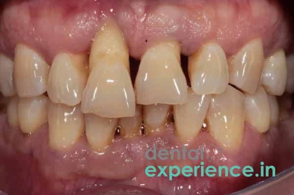
A dental extrusion occurs when a tooth is displaced upward and outward from the socket. The tooth is partially pushed out of its bony pocket. It does not fall out of the mouth, unlike what happens when a tooth is avulsed. Instead, it sticks up from the gum line. Extruded teeth appear longer than their counterparts because they are displaced along the length of the tooth. This happens due to trauma to the periodontal tissues — the structures that anchor the tooth to the jawbone. The periodontal ligament fibers, which provide a cushioning effect around the tooth like a springboard, become stretched or broken. Inside the tooth, the nerves and blood vessels enter through a small hole called the apical foramen located at the bottom of the root. These are usually cut off or pinched as well, affecting circulation to the tooth’s interior pulp.
Definition: Partial displacement of a tooth from its socket caused by injury to the periodontal ligament.
Urgent Indications for Treatment: There are specific indicators that show an urgent need for treatment: an elongated appearance; increased movement of the tooth; hemorrhaging from the gums; extreme sensitivity or discomfort; an open gap in the bite, etc.
Treatments: Treatment options include stabilizing the tooth with a splint; performing endodontic therapy (root canal); restoring the tooth with a crown or implant; removing severely damaged teeth.
Risk Factors: Pulp death, continued resorption of roots, and localized bone loss around the tooth are some potential risks associated with dental extrusions.
Visual Appearance: All patients who experience a dental extrusion report seeing one of their teeth protruding below the level of their other teeth. Their teeth appear visually elongated.
Extreme Mobility: Patients report extreme looseness or mobility when they touch their teeth with their tongues or lips. Patients describe feeling the displaced tooth hit prematurely against opposing teeth, making normal jaw closure impossible.
Occlusal Interference: A common symptom among many patients who have had a dental extrusion is occlusal interference. They cannot close their mouths. Their teeth hit before they can even start closing their mouths. Because of this, most patients have difficulty eating or drinking anything because it creates extreme discomfort.
Gingival Bleeding & Swelling: When patients have an extruded tooth, there is bleeding from the gums. Many patients report constant or recurring bleeding. This is due to the fact that the capillaries surrounding the tooth have been damaged.
Altered Sensation: Due to the exposure of dentin and damaged nerve endings, patients report an altered sensation. Some report no sensation at all while others report extreme pain or soreness from previously minor sensations such as cold air.
Warning signs to look for immediately after a dental extrusion indicate serious damage and possible complications:
Blackening: If the tooth begins to darken quickly (within a couple of hours), it is likely due to pulp necrosis and should be treated immediately. Rapid necrosis of the pulp is indicated by rapid blackening or purpling.
Swelling Beyond Gum Line: If swelling develops beyond the gum line into areas outside the immediate vicinity of the tooth, i.e., into areas of facial spaces such as cheeks, submandibular area, etc., it could be indicative of an advancing infection.
Aspiration Risk: If the tooth is so loose that it may dislodge while swallowing, it becomes an immediate aspiration hazard (dangerous to breathe in). Therefore, if either of these conditions occur, seek emergency help right away.
Possible causes of dental extrusions include:
Traffic accidents: In Kerala, an extremely common cause of dental extrusions is motorcycle collisions on twisting rural highway roads where motorcyclists collide with motorcycle handlebars or roadside objects without protective headgear.
Industrial or agricultural mishaps: Agricultural or industrial falls onto heavy equipment or large branches may produce enough force to dislodge teeth from sockets.
Biting down on unusual objects: Biting down on something that has unusual texture or hardness and slipping off, creating uneven forces on a structurally weak tooth may lead to tooth displacement from its socket.
Contributing factors that make a person more susceptible to tooth displacement include:
Periodontitis: Chronic gum disease destroys the alveolar bone and gradually thins out the periodontal ligament fibers. As a result, teeth with periodontitis lose much of their structural support and are far more prone to dislodgment than otherwise healthy teeth.
Maxillary Protrusion: The more pronounced a patient's upper front teeth protrude (so-called "buck" teeth), the higher their chance of suffering from displacement injuries to those teeth. Maxillary prominence increases the likelihood of direct trauma striking the unprotected teeth.
Short Roots: Teeth with inherently short or thin roots have fewer surface areas anchored deep within the alveolar bone. Consequently, these roots are relatively easy to dislodge from their sockets upon encountering sufficient force.
Lack of Mouthguards: Involvement in contact sports without wearing custom-made athletic mouth protectors greatly increases risk of trauma to teeth and surrounding tissues.
Remember do not try to force/pull your tooth back into place with your fingers since you could further compress/damage what little remains of your blood vessels!
Alignment Problems
└── Look at your smile line. Does one tooth hang lower than all your other teeth?
└── Take a closer look for any visible bands of darkened roots near your gum lines.
└── Try to slowly close your mouth together. Are your teeth coming together evenly?
└── If your loose tooth bumps into another tooth first and prevents your teeth from closing properly together then you have experienced an extruded tooth.
If there is active bleeding from between your gums and your tooth then place some moist cotton gauze lightly against this area and hold it firmly for a moment until you arrive at the dentist office.
Do not touch your loose tooth until you arrive at the dentist office
Dental professionals use a very detailed process to determine whether a tooth has been displaced. A thorough clinical evaluation begins with a soft examination of the affected area, followed by determining how much the tooth can be moved horizontally by applying pressure to the handle of each instrument being used.
|
Diagnostic Steps |
Tools/Methods |
Purpose |
|
Radiographic Assessment |
X-rays (periapicals) or CBCT images |
To see the expanded interdental spaces that appear as a result of the widening of the PDL and to verify whether there is a horizontal fracture to the root. |
|
Vitality Assessment |
Cold Spray Thermal Test (thermal testing), or Electric Pulp Tester (EPT) |
To see if the internal nerve bundles are sending signals to the patient due to electrical stimulation, or thermal sensation. |
|
Occlusion Analysis |
Articulating Paper Test |
To find the areas of high contact in your bite where you are placing harmful force on the tooth that has been damaged. |
Determines the pathway of management for an extruded tooth based upon the level of structural damage caused to the displacement of the tooth:
Grade I (Minimal): Displacement less than 2 mm. The tooth is somewhat loose, however, all of the supporting alveolar bone remains structurally intact.
Grade II (Moderate): Between 2mm and 4mm of displacement. There is great movement of the tooth both vertically and laterally. Part of the support structure of the tooth, i.e., the periodontal attachments have been partially torn.
Grade III (Extensive): More than 4mm of displacement. The tooth is loose enough so that it may hang by just a few strands of connective tissue. It is also common for labial or palatal portions of alveolar bone plates to be fractured.
Surgical restoration of an extruded tooth involves multiple phases. Initially, a dentist will relocate the tooth back to its anatomically correct position under topical anesthesia.
After relocation, a flexible splint made of composite resin and orthodontic wire is applied. An orthodontic wire, or ribbon made from fiberglass, is attached to several teeth adjacent to and above or below the injured tooth. To hold everything together, this is then bonded to those teeth using flowable composite resins. The flexibility of this type of splint allows for minute movements as part of normal function; these encourage fibroblasts to repair and form new connections to attach the periodontal ligament fibers back onto the roots. This healing process takes nearly 2 weeks.
As extrusion injuries typically sever the delicate blood supply of adult permanent teeth, root canal treatment (endodontic therapy) is indicated within 7 to 14 days of the injury for teeth with fully closed, mature roots to prevent internal complications. For immature permanent teeth with open root apices, the pulp has a chance at spontaneous blood supply revascularization and is monitored closely before deciding on endodontic therapy. After removing dead internal pulpal tissue and cleaning/sanitizing the root canals using calcium hydroxide medicaments or bioceramic materials, the dentist seals off the pulp chamber with gutta percha to prevent an abscess forming around the tooth. Following verification of internal healing processes, a custom-made zirconium or porcelain fused-to-metal (PFM) crown reinforces structural integrity of the tooth.
The cost involved in saving an extruded tooth in Kerala is dependent on the extent of the trauma experienced and on the restorative material chosen.
Emergency stabilization using local anesthesia and wire splints generally falls in the range of ₹1500 to ₹4000 across private clinics located in urban centers such as Kochi, Trivandrum, and Kozhikode.
If endodontic intervention is necessary, a rotary root canal treatment adds approximately ₹3,500 to ₹7,000 to this base cost.
Finally, restoring structural integrity using a dental crown is solely dependent on the material used for construction: a standard PFM crown averages ₹3000-₹6000 while highly aesthetically pleasing monolithic Zirconia crowns average ₹8000-₹18000.
Public Sector Government Medical and Dental Colleges provide all these services at highly discounted prices although waiting periods for non-emergency phases may vary greatly.
A general dental practitioner can handle the first emergency repositioning and simple splinting. A group of dental specialists will need to be involved in treating a more serious case.
Endodontists: The root canal specialists are key when dealing with a small, calcified, or distorted root canal to preserve the tooth from becoming discolored or infected.
Periodontists: The gum and bone specialists need to be involved in a situation where there is a lot of damage to the periodontal ligament or where surgical treatment of a fractured alveolar bone socket is required.
Prosthodontists: The reconstructive specialists will create and apply advanced crowns or bridges if the structure of the natural tooth needs extensive repair, both cosmetically and functionally.
Time is everything when it comes to preserving a natural tooth as the result of an extruded tooth. In many cases, the most beneficial way to keep a tooth alive is to put it back into its bony socket before 2 to 4 hours pass. The closer the time frame is to 0, the better chance the tooth has of keeping its PDL intact and avoiding external aggressive root resorption.
If treatment is done 24 to 48 hours late, the blood clotting that takes place at the bottom of the socket forms and organizes so well that getting the tooth manually placed in the correct location becomes very difficult. At times, removal of the tooth is the best option.
Will an extruded tooth heal itself back to normal even if I do nothing?
No. An extruded tooth can never find its own way back into its bony socket. After being left unattended, the tooth will grow permanently in its elongated position. That creates problems for your bite. It creates chronic root infections. And it will cause the tooth to come loose because of lack of bone support around the tooth.
Will my tooth turn black after it has been relocated, and splinted?
A mature, permanent tooth can’t get blood flow after it has been extruded. Without having a timely root canal treatment, the dead material inside the pulp chamber will break down. The dead material inside the pulp chamber will release iron pigments which stain the internal dentin and make the tooth look gray or black.
How much pain am I going to experience when having an extruded tooth put back into its proper socket?
The whole process of repositioning and splinting is done using local nerve blocks or infiltration anesthesia. This totally numbs the injured area so that you just feel slight pressure but not sharp pain when they place the tooth.
Clinically, management of an extrusive luxation requires prompt action to conserve attachment apparatus. The previous method of rigid immobilization of an injured tooth was replaced by a temporary fixation for approximately 2 weeks. By doing so, we stimulate differentiation of mesenchymal stem cells in periodontal ligaments and avoid ankylosis (fusion of roots to jawbone).
Early endodontic extirpation of necrotic pulp tissues within 2 weeks is also important to avoid complications from post-traumatic internal infection. Long term clinical follow up at 4 weeks, 3 months, 6 months and 1 year is mandatory to ensure structural stability and monitor for silent root resorption.














.jpg)
.jpg)

.jpg)
.jpg)

.jpg)
.jpg)
.jpg)
.jpg)
.jpg)
.jpg)
.jpg)
.jpg)