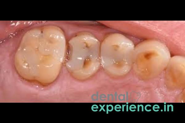
A fractured filling is defined as a dental restoration that has either cracked, fractured, or structurally failed. As a re-do case in both operative and restorative dentistry, a fractured filling can occur when the dental material (composite resin, amalgam, or glass ionomer) loses its seal between the dental material and the surrounding enamel (the hard outer shell of the tooth). Once the dental material and the surrounding enamel lose their structural bond, the underlying dentin—the layer of the tooth containing many microscopic tubules—is exposed. This provides a pathway for bacteria to travel up the dentinal tubules to reach the pulp chamber housing the tooth's nervous tissue.
Clinical Definition: Failure of structural integrity in an existing composite, amalgam, or GIC restoration.
Signs That Require Urgent Attention: Sudden onset of throbbing pain, localized swelling of the gums adjacent to the fractured restoration, sharp pain while chewing, visible structural deterioration allowing access to a darkened cavity.
Treatment Options: Resin composite bonding, porcelain inlays/onlays, or full coverage crowns in combination with endodontic therapy if the fracture extends into the pulp chamber.
Sensory Differences: A sharp, jagged edge located by running the tongue across the tooth surface, sensation that the tooth "breaks apart" or is "hollowed out" during eating.
Thermal Sensitivity: Sharp, transient pain generated when drinking a hot beverage such as chai tea, or an icy drink.
Pain: A sharp flash of pain upon releasing biting pressure indicates that the physical tooth structure itself may be cracked (Cracked Tooth Syndrome), which often co-occurs with or mimics a fractured filling.
Food Dislodgement: Debris from food sources (particularly fibrous local food), consistently becomes lodged within the newly created structural gap.
While a minor crack may be tolerated for a couple of days, there are certain clinical warning signs that indicate prompt intervention is warranted to avoid necrosis (death) of deep tissues:
Continuous Throbbing Pain: Persistent, unprovoked throbbing pain that continues unabated without an apparent cause of provocation, suggesting pulp inflammation.
Night Time Pain: Nocturnal pain increases significantly when lying flat (in a supine position) because the loss of gravity increases blood flow and pressure to the head, compressing the inflamed nerves within the rigid walls of the tooth.
Localized Gum Swelling: Small pimple-like swelling (abscess) on gum adjacent to fractured tooth.
Bad Taste Or Foul Smell: Persistent bitter taste or foul smell originating from a region of the tooth with fractured filling indicates presence of large amounts of anaerobic bacteria fermenting in the space beneath loose filling.
There are no circumstances under which a filling will fail unless subjected to some type of stress or strain:
Mechanical Stress: Consuming excessive quantities of hard foods such as fried banana chips, unpopped corn kernels, etc., or chewing directly on hard foods.
Fatigue: Older dental amalgams undergo 'creep,' a process characterized by a gradual deformation of the metallic structure due to prolonged exposure to masticatory forces. Over a period of 10 to 15 years, creep results in numerous micro-cracks throughout the entire filling structure.
Secondary Caries: Bacterial migration through microscopic crevices between dental material and surrounding enamel produces caries at the base of the existing filling. Gradual dissolution of tooth structure by bacteria eventually results in collapse of the filling.
Habitual Parafunction: Frequent nighttime grinding (bruxism) subjects teeth to extremely high force. These forces shear off portions of previously placed restorations.
Several risk factors exist that compromise structural integrity and increase susceptibility to fracture:
History Of Large Cavity — Cavities that were so extensive that more than 50% of the original tooth structure was lost resulting in weakened supporting enamel walls.
Acidic Diet: Excessive consumption of acidic foods or beverages containing strong dietary acids (like citric or phosphoric acid) accelerates the degradation of bonding interfaces between composite resins and tooth surfaces.
Dry Mouth: Saliva contains buffering minerals that help protect against acid erosion of tooth structure. Lack of saliva (xerostomia) rapidly facilitates development of secondary caries along borders of restorations.
Age of Filling: With passage of time, all fillings degrade structurally and chemically.
If a patient suspects that he/she has a fractured filling, the following safe examination should be performed visually in front of a light source and tactilely with a gauze sponge:
Visual Examination: Gently dry area with gauze sponge. Visual inspection for black lines radiating from borders of existing filling, or clearly visible separation between tooth structure and existing filling material.
Tactile Examination: Run tip of tongue lightly over tooth surface to identify new sharp edges or cratering within area of suspected fracture. Avoid inserting metal toothpicks, safety pins, or fingernails into areas suspected of having been compromised. Such actions could create additional weakness within fragile enamel or push bacteria further into the pulp chamber.
Pressure Bite Test: Apply gentle pressure on the tooth with a clean cotton roll. Record presence or absence of pain during pressure phase and/or relief phase.
Accurate diagnosis involves clinical evaluation utilizing precision instruments:
Visual/Tactile Evaluation: Dentist examines tooth visually/tactically to determine presence of catching, softness, or gaps at borders between existing filling and surrounding tooth structure.
Radiographic Evaluation: Digital intraoral periapical (IOPA) radiographs assess depth of fracture and potential presence of secondary caries developing below remaining filling material.
Evaluation Of Surrounding Periodontal Status: Periodontal probing evaluates extent of involvement of periodontium adjacent to fractured area.
Evaluation Of Tooth Vitality: Utilizing thermal/electric pulp tests assesses vitality/potential for nerve infection within tooth structure.
Restorative failures are graded into distinct clinical levels to determine the ideal treatment pathway:
|
Severity Level |
Structural Damage |
Symptoms |
Treatment Approach |
|
Grade 1: Superficial |
Minor marginal chipping of the filling material, the enamel remains intact. |
Mild roughness, no pain or thermal sensitivity. |
Smooth edges or minor composite resurfacing. |
|
Grade 2: Moderate |
Structural loss of the filling exposing the underlying dentin layer. |
Short, sharp sensitivity to hot, cold, or sweet inputs. |
Complete replacement with a direct composite or durable inlay. |
|
Grade 3: Advanced |
Extensive fracture reaching the pulp chamber or extending below the gumline. |
Severe, throbbing pain, lingering thermal sensitivity, tooth mobility. |
Root canal therapy followed by a full-coverage dental crown. |
Options available to modern dentistry include highly durable methods to restore a fractured filling:
Composite Resin Bonding: Minor-to-moderate fractures repaired by removing old material and replacing with nano-hybrid composite resins matching color of adjacent tooth structure exactly.
Porcelain Inlays/Onlays: Moderate-to-extensive fractures repaired by removal of portion of existing tooth structure and fabrication of porcelain/zirconia insert by lab technician for insertion onto prepared tooth.
Full-Coverage Crown Fabricated From Monolithic Zirconia: Extensive fractures repaired by covering remainder of tooth structure with monolithic zirconia crown providing high degree of resistance to fracturing.
Glass Ionomer Cement (GIC): Biocompatible material used as subbase to provide protection against structural weakening, or as therapeutic temporary material providing fluoride release to gradually remineralize weakened dentine matrix.
Estimated costs associated with treatment vary greatly depending upon choice of material, clinical complexity and regional pricing:
Direct Composite Fillings: Cost ranges from ₹1200/-to ₹3500/-per tooth based on size of initial cavity.
Root Canal Therapy (if required): Estimated costs range from ₹3500/-to ₹7000/-based on location of tooth within mouth.
Ceramic/Zirconia Crowns: Estimated costs range from ₹5000/-to ₹15000/-+ per unit.
Basic restorations are sometimes partially covered under state-sponsored medical plans or private insurance plans offered by hospitals. However, most premium quality ceramic/zirconia materials are typically non-covered expenses paid by individual patients.
Choosing a correct dental specialty ensures that successful long term outcome is achieved:
General Dental Practitioner: Appropriate general practitioner for evaluating problems related to fractured fillings, performing routine direct composite/GIC restorations, repairing uncomplicated fractured fillings.
Endodontist: Specialist called upon when bacterial infections spread from the pulp chamber down into the root canals of the tooth; required when severe inflammatory responses develop.
Prosthodontist: Specialist in restoring form/function/replacing missing teeth, called upon for complex restorative needs including multiple porcelain onlays/full-mouth reconstruction/aesthetic crowns.
You can prevent a small crack from becoming a large extraction if you act quickly:
Immediate (within 2-4 hours): If you are experiencing facial swelling, continuous throbbing pain that is unresponsive to medication, or active bleeding from the surrounding gum tissue.
Urgent (within 24-48 hours): If you experience severe toothache while eating or extreme sensitivity to cold temperatures due to exposed dentin.
Elective (within 7-10 days): The tooth has a small, smooth chip, no pain and no sensitivity, no functional problems.
What will happen to my tooth if I don't treat a broken filling for a few months?
If you don't take care of a broken filling immediately, oral bacteria will begin to colonize the exposed dentin tubules. This will eventually lead to deep secondary decay. As a result, you'll likely develop a severe infection within the tooth's nerves. In turn, you could create a massive abscess that would require either a root canal procedure or removal of the affected tooth.
Will a broken filling give me a bad taste in my mouth or make my breath smell terrible?
Yes. Bacteria grow rapidly in the microscopically-sized crevices created when a filling becomes dislodged. These areas become prime locations for the bacteria to ferment food particles and release hydrogen sulfide gas, which is responsible for many of the foul odors associated with halitosis.
Are over-the-counter temporary dental cements available at pharmacies safe to use?
Only for short periods of time (up to 24 to 48 hours), these kits are perfectly safe to temporarily cover sharp edges. However, they do nothing to eliminate bacteria or provide a long-term seal of the tooth. Using one of these products for longer than intended will merely trap all of the decay under the old filling, and accelerate the process of degrading your tooth structure.
The key point here from a clinical standpoint is that a fractured filling should not be considered as simply a failure of material properties, but rather as a sign that the physical environment of the mouth has been altered. This alteration may arise from secondary decay destroying the enamel or possibly as a result of unknown nocturnal bruxism. Prompt attention to restore the structural integrity of the involved tooth is essential. By doing so, you preserve natural tooth morphology, prevent excessive gingival recession, and avoid invasive endodontic therapies entirely.














.jpg)
.jpg)

.jpg)
.jpg)

.jpg)
.jpg)
.jpg)
.jpg)
.jpg)
.jpg)
.jpg)
.jpg)