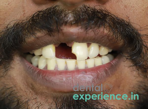
Missing a single tooth, a condition defined as localized partial edentulism, affects much more than aesthetics. When a tooth is lost, there is a cascade effect of structural failure throughout the entire masticatory apparatus. Each tooth functions as a structural component to maintain the balance of the entire apparatus. Teeth functionally support each other and maintain occlusion with their counterparts in the opposing arch.
Loss of a tooth causes a series of structural changes. Without chewing pressure to help maintain the density of the jawbone, the bone tissue will shrink permanently (alveolar bone resorption). As the teeth lose their lateral support, they begin to migrate or tilt towards the area of the missing tooth (pathological migration). On the opposite arch, the tooth loses its biting partner, causing it to grow beyond its socket boundaries (supra-eruption).
Definition: Loss of a single tooth from the dental arch.
Structural Complications: Primary complication includes rapid and progressive loss of alveolar ridge bone. Secondary complications include shifting, tilting and misalignment of adjacent and opposite teeth.
Indications for Urgent Interventions: Patients should seek emergency care immediately if they experience acute localized pain, sudden swelling or rapid noticeable changes in jaw alignment.
Standard Treatment Option: Gold standard treatment option includes placing a biocompatible titanium dental implant that mimics the natural root function.
Treatment Timeframe: Best results are obtained by replacing the tooth within three to six months. Longer delays may result in irreversible bone loss requiring costly bone grafting procedures later.
The most notable symptom associated with losing a tooth is the visual appearance of a large cavity in the dental arch. Once the tooth is lost, there is a significant change in oral sensory awareness. Speech patterns and tactile sensation will change.
Patient reports persistently moving their tongue into the space left after the tooth was extracted.
Increased Masticatory Effort: Difficulty in breaking down solid foods to eat; forces patients to transfer food to the other side of the mouth to chew.
Jaw Muscle Strain: Patient experiences dull aching, stiffness or localized muscle strain in the jaw and surrounding area resulting from inefficient chewing.
Food Entrapment: Constant entrapment of food particles in the narrow, uneven spaces developing as adjacent teeth begin to tilt into the space left by the missing tooth.
Halitosis/Lost Taste: Chronic halitosis (bad breath) and/or unpleasant tastes develop as the result of plaque biofilm accumulation in difficult-to-clean locations.
Schedule an immediate appointment with your clinician if you notice any of the following warning signs:
Acute Inflammation Signs: Sudden sharp pain, swelling in the gingival tissues surrounding the empty socket or redness.
Suppurative Drainage/Pus: Presence of any foul tasting discharge from the empty socket indicates active bone infection.
Loosening/Mobility/Increased Sensitivity of Adjacent Teeth: Immediate mobility or increased sensitivity of teeth bordering the defect.
Occlusal Changes/Shifting: Sudden changes in occlusion including clashing front teeth or loud audible clicking sounds when opening/closing jaws.
Single tooth loss occurs as a direct consequence of mechanical/biological/traumatic injury affecting the supporting structures holding the tooth in place.
Advanced Untreated Chronic Periodontitis: Systemic bacterial attack destroys PDL and supporting bony structure until tooth falls out or must be removed.
Deep Carious Lesions: Aggressive dental caries leading to complete invasion of the pulp chamber through all layers of enamel/dentin/tooth structure destroying structural integrity of the tooth.
Traumatic Injury: Forces applied mechanically during sports related injuries, fall, vehicle accident etc., causing dislodgement from socket of tooth.
Mechanical/Biological/Dietary Hazard Trauma: Biting into hard objects found in local cuisine (e.g., rice stones, unrefined jaggery, or brittle banana chips) frequently causes severe structural fractures. If these fractures extend vertically down the root, the tooth becomes unrestorable and must be extracted, creating the single tooth gap.
There are several underlying systemic conditions/lifestyle factors that contribute to weakened dental structures and accelerated tooth loss.
Uncontrolled Diabetes Mellitus: Elevated levels of blood glucose damage microvasculature impairing immune response and accelerating aggressive bone loss around teeth; extremely common among population in Kerala.
Smoking/Tobacco Consumption/Betel Nut Chewing: Decreases blood circulation to gums masking early signs of infections and rapidly increasing rate of destruction of jawbones.
Poor Home Oral Hygiene Habits/Habits: Failure to properly clean/floss surfaces creates environments for aggressive bacteria to form sticky plaque biofilms that then calcify into resistant tartar deposits creating chronic sites for further destruction.
Generalized Systemic Bone Loss/Osteoporosis: Reduction in overall bone density found in aging populations contributes to general thinning/weakening of maxillary/mandibular bones and potentially enhances rates of alveolar ridge bone thinning.
Using a well-lighted mirror, perform this non-touch self-assessment examination to preliminarily assess your missing tooth space:
Inspect Gingiva Surrounding Gap: Inspect color/pinkness/flattened shape/gum recession around missing tooth space. Healthy gums should look pink and smooth; inflamed/swollen/red gums suggest infection.
Measure/Assess Adjacent Alignment: Assess positioning/vertical orientation/movement of adjacent teeth to gap. Should be straight/equidistant from centerline; movement/tipping indicates pathology.
Assess Opposition Arch Position: Assess positioning/movement/tilt of opposition arch tooth. Should sit parallel/same plane as adjacent teeth; upward drift/migration suggests supra eruption.
Evaluate Jaw Function/Occlusion/Bite: Close jaws gently. Evaluate for even/joint/uncomfortable sliding motion or pain.
A complete assessment of necessary restorative planning necessitates thorough clinical evaluation utilizing diagnostic equipment to document anatomical relationship between bone and nerves around missing tooth gap.
Steps included in a practitioner diagnosis:
Clinical Periodontal Probing: Dentist evaluates health status of gums surrounding adjacent teeth using a measuring tool (periodontal probe).
Intraoral Periapical (IOPA) Radiographs/Xrays: Provide high-quality 2-dimensional digital image representations of surrounding adjacent root structures and remaining bone height.
Cone Beam Computed Tomography (CBCT): Measures actual dimensions/widest points/density/volume of jawbone required for safe placement of implant.
Pulp Vitality Testing: Uses gentle heat/electric stimulation to verify that nerves of adjacent "anchor" teeth are fully intact/healthy
The clinical severity of a single missing tooth is graded based on how long the tooth has been missing, the amount of bone loss, and the movement of surrounding teeth.
|
Severity Level |
Clinical Presentation |
Functional Impact |
|
Grade 1: Mild |
Recent extraction site; healthy bone volume; adjacent teeth perfectly upright. |
Minimal; minor speech or chewing adjustments needed. |
|
Grade 2: Moderate |
Early bone height reduction; neighboring teeth beginning to tilt; slight over-eruption opposite. |
Minor bite imbalances; frequent food trapping in new gaps. |
|
Grade 3: Severe |
Advanced bone resorption; severe tilting of neighbors; opposite tooth heavily over-erupted. |
Major chewing dysfunction; structural jaw joint strain. |
Dentistry today provides three very successful methods for replacing one missing tooth. Each method depends upon the patient's individual dental needs, health considerations and budget constraints.
Single tooth dental implants: Single tooth implants are considered to be the "gold standard" of dental care. In this method, a titanium implant is inserted directly into the jawbone through surgery. After three to six months, a process known as osseointegration will occur; during this time the bone will integrate itself with the metal implant. Once integrated, a customized ceramic crown is attached to the implant, allowing the patient to eat normally and preventing any contact with other teeth.
Fixed dental bridges: Fixed dental bridges consist of an abutment area on each side of a missing tooth. These abutments consist of an anchor pillar made of trimmed-down tooth structure on the sides of the two healthy teeth next to the missing tooth. A pontic tooth is suspended across the gap created by the missing tooth by attaching a chain of three connected prosthetic crowns.
Removable partial denture (RPD): A removable denture consists of an acrylic or metal frame that holds an artificial tooth. It is clipped onto remaining teeth. Although it is inexpensive, a RPD must be removed every night and does not slow or stop bone loss around the missing tooth.
Kerala-based dental clinics use standard international systems for dental implants. Depending on the material(s) used and the premium level of the system chosen by the clinic, prices vary from ₹25,000 to ₹45,000 per unit. Bone grafts needed prior to placing a dental implant can increase the price to anywhere between ₹50,000 to ₹75,000.
Prices for a traditional three-unit porcelain fused to metal (PFM) fixed bridge are about ₹9,000 to ₹18,000. Prices for high strength Zirconia Fixed Bridges are between ₹20,000 to ₹40,000.
Simple acrylic partial RPDs are extremely low-cost and generally fall in a price range of ₹2,000 to ₹5,000.
As most of India's private insurance companies classify single tooth restoration as cosmetic treatment and therefore do not cover them under medical/dental insurance, these types of treatment must be paid for out of pocket. However many large dental practices in cities such as Kochi, Trivandrum and Kozhikode offer flexible 0% interest monthly payment plans (EMI) to make it easier for patients to afford this type of treatment.
Prosthodontists: Your Prosthodontist is a specialist whose focus is exclusively on creating and providing functional and aesthetic artificial teeth replacements including crowns, bridges and implant restorations.
Oral & maxillofacial surgeons/Periodontists: If your case requires an extensive amount of work such as a complicated placement of a dental implant or an advanced bone grafting procedure then you will need to see an oral & maxillofacial surgeon/periodontist. They are both highly trained surgeons specializing in surgical procedures related to the oral cavity.
Endodontist: An endodontist is a specialist who specializes in root canals. They will evaluate if the adjacent teeth need to be treated endodontically and confirm that the roots and internal canals are healthy enough to serve as long-term foundations
Depending upon how quickly you receive treatment after losing a tooth there are different timelines associated with receiving treatment.
Traumatic Avulsion – time sensitive (1 hour): In the event that a healthy natural tooth is completely avulsed due to an injury, keeping the tooth moist in cold milk, Hank’s Balanced Salt Solution (HBSS), or the patient's own saliva while rushing to a local dentist may allow you to save some or all of your natural root ligaments. Attempting to re-implant it immediately with your local dentist can potentially restore your tooth.
Elective restoration – not as time sensitive (3–6 Month Window): When a tooth is lost due to decay or gum disease it is recommended to establish a permanent solution within six months. Allowing more than six months to pass creates opportunities for bone loss to occur. Bone loss makes future implant placement far more difficult and expensive.
Do I really need to replace my missing upper molar since I don't show it when I smile?
Yes – since your upper molars carry the bulk of your mastication forces, leaving an upper molar gap will cause adjacent teeth to tilt, rotate or shift forward. The result will be malalignment of your occlusion (the way your upper and lower jaws meet), increased wear on your front teeth and tmj disorders.
How long does the entire single tooth dental implant process take from beginning to end?
Generally, the complete single tooth dental implant process can take up to six months from start to finish. This extended timeframe allows adequate opportunity for the bone in your jaw to fuse tightly around the titanium implant surface prior to loading with chewing forces.
Can an elderly person with type 2 diabetes safely receive a dental implant?
Yes – as long as you maintain good control over your type 2 diabetes through regular medication therapy, an elderly person can safely receive a dental implant. Elderly patients with well-controlled type 2 diabetes experience highly successful outcomes comparable to non-diabetics, though clinicians may monitor them closely as systemic healing pathways can occasionally be slightly prolonged.
Replacing a single missing tooth promptly is an essential preventive measure for your long-term health. When a gap is left unaddressed, the entire mouth undergoes a negative structural shift that compromises chewing and strains the jaw. Modern implant dentistry allows us to replicate natural anatomy perfectly, protecting neighboring teeth and keeping the jawbone strong. Taking action early ensures predictable, long-lasting outcomes and prevents complex bite alignment problems down the road.














.jpg)
.jpg)

.jpg)
.jpg)

.jpg)
.jpg)
.jpg)
.jpg)
.jpg)
.jpg)
.jpg)
.jpg)