
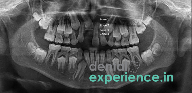
An unerupted tooth occurs when a tooth develops but never breaks through the gums to emerge in the mouth. Clinically, an unerupted tooth is classified as an anomaly related to tooth positioning due to the fact that it causes a misaligned bite and/or an aesthetically unacceptable smile when a prominent front tooth is involved such as the upper central incisors or maxillary canine.
As a result of being trapped beneath the gum and bone, a tooth remains buried in the alveolar bone and covered by a protective dental follicle. As a direct result of the tooth not emerging, it also displaces adjacent teeth which may shift toward the gap. Ultimately, this will result in both uneven smiling and severely displaced bites.
Definition: An unerupted tooth is any tooth that remains beneath the gum line, which is normal during early development. However, it becomes classified as an impacted tooth when it is pathologically blocked from emerging by a physical barrier (like bone or another tooth) or because it has drifted completely off its normal eruption path past its expected chronological timeline.
Emergency Signs: Rapidly spreading facial swelling, severe throbbing pain that disrupts sleep, or swelling accompanied by a fever.
Treatment Options: Surgical access for orthodontic movement of the impacted tooth (tooth pulled down by means of braces); surgical extraction followed by placement of dental implants; or space maintainer options including cosmetic bridges.
Individuals who have an unerupted tooth will frequently exhibit notable physical and tactile symptoms in their mouths. Visually, the most apparent symptom is a large gap where a permanent adult tooth should have appeared. Due to the fact that all of the teeth on the opposite side of the mouth appear fully erupted, this creates visible asymmetry that disrupts the natural smile aesthetics and dental arch balance.
Physically, patients may detect a hard smooth mass located underneath the gums where the crown of the trapped tooth is pushing against the bone. Neighboring teeth may begin to tip or drift into the empty space creating changes in how the top and bottom teeth meet. Eventually, this tipping can create chronic fatigue in your jaw muscles.
While it appears to be a merely cosmetic issue of having a missing tooth, several clinical warning signs indicate possible deeper soft-tissue damage and/ or impending bone loss requiring urgent treatment:
Asymptomatic Cysts: While dentigerous cysts forming around the crown of a trapped tooth are usually painless and completely asymptomatic in their early stages, they can silently expand and destroy surrounding jawbone. Pain typically only occurs if the cyst becomes acutely infected or leads to a structural bone fracture.
Resorbing Roots: The pressure from the path of the unerupted tooth can trigger cellular activation that causes external root resorption (dissolving) of the roots of adjacent healthy teeth.
Recurring Gingival Inflammation: Ongoing redness, heat, and purulent drainage around the empty space indicates that bacteria have gathered under the gum flap around a partially erupted tooth crown, creating a localized infection known as pericoronitis.
Structural and mechanical barriers that exist within the jaw bone typically contribute to the inability of a tooth to develop properly. Crowding is likely the most common barrier; however, if there is no adequate physical room available for the developing tooth to fit into on the alveolar ridge, then it will become trapped. Retaining an overly matured primary baby tooth also serves as a physical barrier preventing an adult tooth from erupting. Additionally, traumatic injury to facial bones during early childhood (e.g., falling onto a baby tooth forcing it back into the socket) can create irregularities in development of permanent teeth germs resulting in them being directed away from the intended pathway into a trapped state.
There are numerous inherent structural characteristics and behaviors that increase the likelihood that an individual will encounter an unerupted tooth:
Family History: Individuals whose families historically have had narrower dental arches or multiple extra teeth (supernumeraries) are more likely to have impacted teeth.
Bone Disorders: Certain genetic conditions affecting bones (cleidocranial dysplasias) may directly interfere with cellular signaling that facilitates normal tooth eruption processes.
Trauma: Cultural dietary practices in India (e.g., biting on rough traditional foods) prior to weaning can cause fractures in developing primary teeth. If left untreated these fractures can progress into deep scarring of supporting structures leading to locking of an adult tooth in a sub-gum position.
It is relatively safe for individuals to examine their own mouths at home utilizing a mirror, light source and fingers:
Examine Symmetry: Examine your front upper teeth for symmetry. Is there a significant difference between each upper front tooth? Has one of your upper front teeth been fully erupted for longer than 1 year and another is still missing?
Assess the Gum Line: Cleanly wash your hands. Gently touch your index finger along the outer edge of your gum ridge above the area where your tooth is missing. Do you feel a hard, non-painful roundish mass just beneath the gum tissue?
Look For Drift: Examine the teeth immediately adjacent to where your tooth is missing. Have they drifted towards the area where your tooth is missing? Are they beginning to collapse upon themselves like pillars do when collapsing?
A formal clinical diagnosis can only be made after a thorough exam by a licensed dentist utilizing advanced diagnostic equipment. Your dentist will initially make observations about your jaw shape through visual inspection and feeling your bone structure. Then, he/she will perform digital x-rays (intraoral xray or panorex) to determine exactly how far away and structurally damaged your trapped tooth is.
If you need additional information regarding smile symmetry your dentist may utilize Cone Beam Computed Tomography (CBCT). CBCT allows him/her to obtain a three-dimensional image of your tooth showing its relationship with other roots and bone density.
Your dentist will conduct a final thermal vitality test to ensure that your trapped tooth has vital pulpal tissue present.
Dental clinicians categorize an unerupted tooth by degree of difficulty of correction depending upon how far embedded it is in bone and how difficult it will be to move:
|
Severity Degree |
Soft Tissue Cover |
Clinical Manifestations |
Complexity Of Treatment |
|
Low (Simple) |
Sub-Mucosal Impaction |
Crown has cleared the bone, trapped only by thick overlying gum tissue. |
Simple soft-tissue window excision (exposure). |
|
Moderate (Intermediate) |
Incomplete Bony Impaction |
Partially encased in bone, the crown is moderately tipped against neighboring roots. |
Minor bone removal, bracket bonding, and steady orthodontic traction. |
|
High (Difficult) |
Total Bony Impaction |
Completely embedded deep in the bone, oriented horizontally or entirely inverted (upside-down). |
Complex surgical exposure or therapeutic surgical extraction. |
Treating an unerupted tooth to restore proper dental alignment and smile symmetry requires a coordinated plan to safely get the impacted tooth into alignment:
Surgical Exposure & Orthodontic Movement: Oral surgeons expose the impacted crown surgically and attach small orthodontic brackets to each of the impacted and adjacent erupted teeth. Through gentle, consistent force applied by an orthodontist utilizing thin gold wires connected to conventional braces, they gradually pull the impacted tooth downward over many months.
Surgical Removal & Replacement With Dental Implants: If an impacted tooth becomes ankylosed (fused) to the bone, it can not be moved. An oral surgeon extracts it and places a graft in the ridge to maintain it, then later inserts a titanium dental implant covered with a zirconia crown.
Costs associated with treating an unerupted tooth vary depending upon complexity of case and treatment methods utilized. In larger cities throughout Kerala State such as Kochi, Trivandrum, Kozhikode etc., a standard surgical exposure procedure for an impacted tooth costs anywhere from Rs 6000 - Rs 12000. In addition, if orthodontic movement with fixed appliances are required for corrective purposes, total treatment cost can range from Rs 35000 - Rs 75000 depending upon utilization of metallic vs clear/ceramic brackets.
Conversely, if extraction with dental implant replacement are required as part of the treatment plan, costs for high-quality dental implant systems can range from Rs 30000 - Rs 65000 per tooth. Typically, these treatments are performed on an "out-of-pocket" basis; although some newer private insurance companies provide partial reimbursement for initial surgical removal procedure(s).
In order to achieve successful correction of this problem, a team approach involving specialists from different disciplines is required since general dentists are not trained to treat complex structural malpositions:
Oral /Maxillo Facial Surgeons: These surgeons play a critical role in providing safe access to impacted crowns through minimally invasive techniques or performing complex extractions close to nerve bundles without causing irreversible damage to the same.
Orthodontists: These professionals are responsible for designing unique orthodontic movement strategies designed to track movement of impacted teeth into their natural positions and applying customized forces through specially created braces.
Prosthodontists: Should a tooth be deemed hopeless and not salvageable; prosthodontists design and fabricate aesthetic restorative solutions such as crowns or bridges designed to recreate lost aesthetics of patient's smile.
An unerupted or impacted tooth itself rarely requires emergency care. True emergency signs—such as rapid swelling, fever, or throbbing pain—only happen if a secondary complication develops, such as a severe bacterial infection (pericoronitis) spreading into the face, or if the trapped tooth causes an acute infection in the root of a neighboring healthy tooth.
However, it does require timely intervention before permanent malocclusion occurs. While waiting until age 9 or 10 is acceptable for monitoring upper canines, an adult upper front central incisor should erupt around age 6 to 7. If a front incisor is missing or hasn't emerged within six months of the matching tooth on the other side, an orthodontic evaluation should be scheduled immediately by age 7 or 8 to avoid severe developmental delays.
Delaying treatment until late teens significantly increases the potential for that impacted tooth fusing itself permanently into bone rendering it extremely difficult (if not impossible) to mobilize in later stages of life. Early action reduces discomfort during treatment and reduces risk of long-term harm to smile architecture
Will a tooth that has yet to erupt eventually work its way into place on its own?
A tooth cannot grow into place if it is obstructed by large amounts of bone or if it is going in the wrong direction. A child tooth may "work itself loose" over a period of time due to minor soft tissue blockages; however, all but rarely do adult teeth erupt after they have passed their usual time frame. All adult teeth require some form of professional guidance to move them from beneath the gums.
Is the process of exposing the tooth painful?
The procedure is performed under local anesthesia, which completely blocks pain pathways. However, because local anesthetics do not block pressure receptors (proprioception), you will still feel pushed, pulled, and vibrating sensations during the procedure, though it will not hurt.
How long does it take to get the tooth into the correct position?
From the point in time when your orthodontist attaches the orthodontic brackets and chains, most teeth erupt within 6-12 months. However, there are many factors that influence this amount of time such as the depth that the tooth is located below the gums and/or how much distance the tooth must travel.
When managing an unerupted tooth causing facial and dental aesthetics issues, timing is everything. Modern dental strategies favor early intervention, ideally when the root of the trapped tooth is between half and two-thirds formed, taking advantage of the tooth's natural eruptive force. By combining precise surgical exposure with gentle orthodontic care, we can successfully guide these teeth into place. This approach avoids the need for artificial replacements later in life and helps patients maintain a natural, healthy smile.














.jpg)
.jpg)

.jpg)
.jpg)

.jpg)
.jpg)
.jpg)
.jpg)
.jpg)
.jpg)
.jpg)
.jpg)

