
Unerupted teeth are those that have not erupted to their correct positions on the dental arches in their respective times in childhood. An example of an unerupted tooth would be one which has failed to erupt through the gingiva (gum) and alveolar bone into its correct position. Pediatric dentistry classifies this occurrence as either primary retention, which refers to failure of eruption of a primary or permanent tooth without some type of mechanical barrier obstructing it, or secondary retention, which is impacted due to a mechanical obstruction such as crowding, excess soft tissue around a developing tooth bud, or supernumeraries (extra non-developmental teeth) such as a mesiodens. The undisturbed retained dental follicle from the submerged tooth may eventually lead to ectopically positioned tooth development, possible harm to adjacent roots, or cyst formation deep within the child’s jawbone if left untreated for extended periods of time.
Clinical Definition: An unerupted tooth is one that has been delayed or confined under the gumline longer than usual prior to reaching the typical eruption period.
Urgent Indicators: Signs and symptoms of an urgent concern include abnormal growth in the local area with respect to the gums, significant jaw pain, misalignment of teeth, and a long delay in shedding a baby tooth at the appropriate time.
Commonly Affected Teeth: Upper maxillary central incisors, upper canines, and upper second premolars.
Most Effective Solutions: Orthodontic traction to assist in aligning the tooth, surgical exposure via gingivectomy, space maintainers, and targeted removal.
Parents can identify that a tooth is impacted as it will present itself in the following ways:
Unsymmetrical Pattern of Eruption: The tooth on one side erupts normally, but on the other side, there has been no eruption at all for six months.
Palpable Bulging of the Gums: When you run your finger across the top part of the upper arch (the palatal side) or the outside of the gums, you can feel a firm, rounded lump.
Retained Primary Tooth: If a baby tooth continues to be firmly in place longer than the anticipated time frame for it to fall out.
Misaligned Gaps: Teeth in the area are shifting, slanting, or twisting into the gap left by the non-erupted tooth.
Localized Sensitivity: Your child may express some degree of sensitivity or discomfort when they chew firm food, usually mild.
You should seek immediate dental attention if you observe any of the following in your child:
Infection Signs: Swollen gums around the erupted tooth, pus from the erupted tooth, and/or swelling over where the hidden tooth is located.
Bony Swelling or Cysts: If the jawbone begins to swell or form cysts without explanation.
Adjacent Root Resorption: Resorption of roots of healthy teeth next to the non-erupted tooth, causing pain.
Radiating Pressure or Headaches: Radiation of pressure/pressure-type headache and/or sinuses. This is common when the impacted maxillary canine is high.
Blockage Due to Physical Obstructions: Excessive growth of mucous membrane, mesiodens, or benign odontogenic tumors (such as odontoma) that block normal flow.
Loss of Baby Teeth Early in Life: Premature loss of a primary tooth due to decay or trauma creates additional space for the adjacent developing permanent teeth to move into this newly created space, thus blocking the pathway.
Ankylosis Before Maturity: Fusion of a primary tooth root to the surrounding jawbone while the permanent successor remains trapped within its bony crypt, which prevents the underlying permanent tooth from emerging along its normal path.
Abnormal Position of the Developing Permanent Tooth Seed (Tooth Germ): A portion of the developing permanent tooth has grown in the wrong position (to the roof of the mouth and/or alongside another tooth's root). It should have continued to grow down through its proper bony canal.
Narrow Upper Dental Arch: There simply isn't enough room in the upper jaw to allow all of the developing permanent teeth to emerge properly.
Changes in Diet: Consuming soft and overly processed foods reduces the amount of mastication force on the mandible. This reduced force hinders the development of the mandible, thereby reducing the available space for the developing permanent teeth to erupt.
Family History: Having a family history of impacted canine teeth, delayed dental eruption times, or missing permanent teeth.
Prolonged Orofacial Habits That Narrow the Dental Arch: Sucking thumbs for extended periods of time and/or using a thrusting motion with the tongue to swallow food. Both habits can cause a narrowing of the dental arch.
Chronic Systemic Health Conditions: Chronic systemic health conditions which can cause developmental delays including delayed eruption times for all teeth and/or malpositioning of some or all of the developing permanent teeth: hypothyroidism, hypopituitarism, cleft lip/palate syndromes, cleidocranial dysplasia, and Down syndrome.
Traumatic Injury to the Mandible: Traumatic injury to the mandible during the growth and development stage caused by an object that displaces and scars the surrounding gum tissue, also displacing the developing permanent tooth from its normal bony pathway.
Performing a preliminary, visually safe examination is possible by parents at home:
Clean Hands: Always wash your hands prior to inspecting your child’s mouth.
Mirror & Light: Examine your child with their face toward you while positioned under bright lighting. With the help of a mirror (the smaller the better), examine both sides of each arch. Be sure to count the teeth present. Compare the number of teeth from one side of the upper jaw to the other and do the same comparison for the lower jaw.
Palpate: Using an index finger, palpate along the gum ridge. Check for an area where there may be a firm bump (representing a tooth bulge) or feel for a soft tissue mass.
Test Tooth Mobility: Gently run your index finger over any baby tooth located in a delayed eruption zone. Test whether the tooth feels loosely anchored in its socket, which would indicate it is ready to erupt.
Important Note: Do not force hard pressure onto swelling gum tissue or attempt to remove trapped teeth.
A pediatric dentist uses objective diagnostic procedures when evaluating an unerupted tooth:
Visual Examination and Palpation: Evaluate the overall space available within the arch and assess the spatial relationships of unerupted teeth using intraoral digital palpation. Determine if there are any areas where there may be unerupted teeth that are creating bulges along the gingiva.
Radiographic Evaluation: The use of intraoral digital radiographs (periapical and bitewing) will provide a radiographic representation of the exact location of the unerupted tooth, including the status of the developing root, and surrounding bone and tissue.
Three-Dimensional Imaging (CBCT): Cone Beam Computed Tomography (CBCT) technology provides low-dose three-dimensional images of all dental and facial structures and allows the pediatric dentist to accurately determine the relationship of the impacted tooth/teeth to adjacent roots, nerves, etc.
Vitality Assessment: Assess the vitality (health) of adjacent teeth using laser technology to assure that no damage has occurred as a result of the impaction.
|
Level |
Clinical Stage |
Description |
|
Grade 1 |
Mucosal Retention |
The tooth is fully formed but trapped under thick gum tissue; bone path is clear. |
|
Grade 2 |
Partial Intraosseous Retention |
The tooth is partially blocked by alveolar bone or an adjacent tilted tooth root. |
|
Grade 3 |
Complete Impaction / Ectopic Position |
The tooth is buried deep within the jawbone, severely rotated, or driving into neighboring roots. |
The modern practice of pediatric dentistry concentrates on maintaining natural teeth utilizing non-surgical and minimally invasive techniques:
Laser-Assisted Surgical Exposure: The diode laser precisely removes the overlying gum tissue with minimal bleeding, cleanly exposing the crown of the impacted tooth.
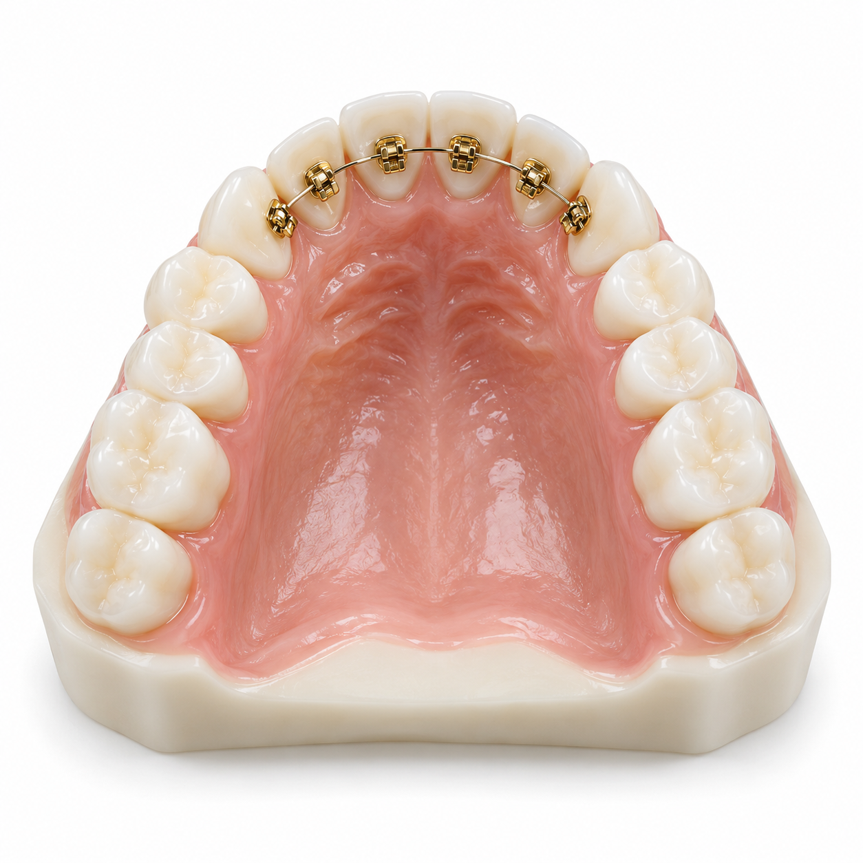
Orthodontic Traction: After exposing the impacted tooth, an orthodontic bracket can be applied. Light gold chain tension can then be gradually increased in a series of monthly visits to slowly move the impacted tooth into its final location.
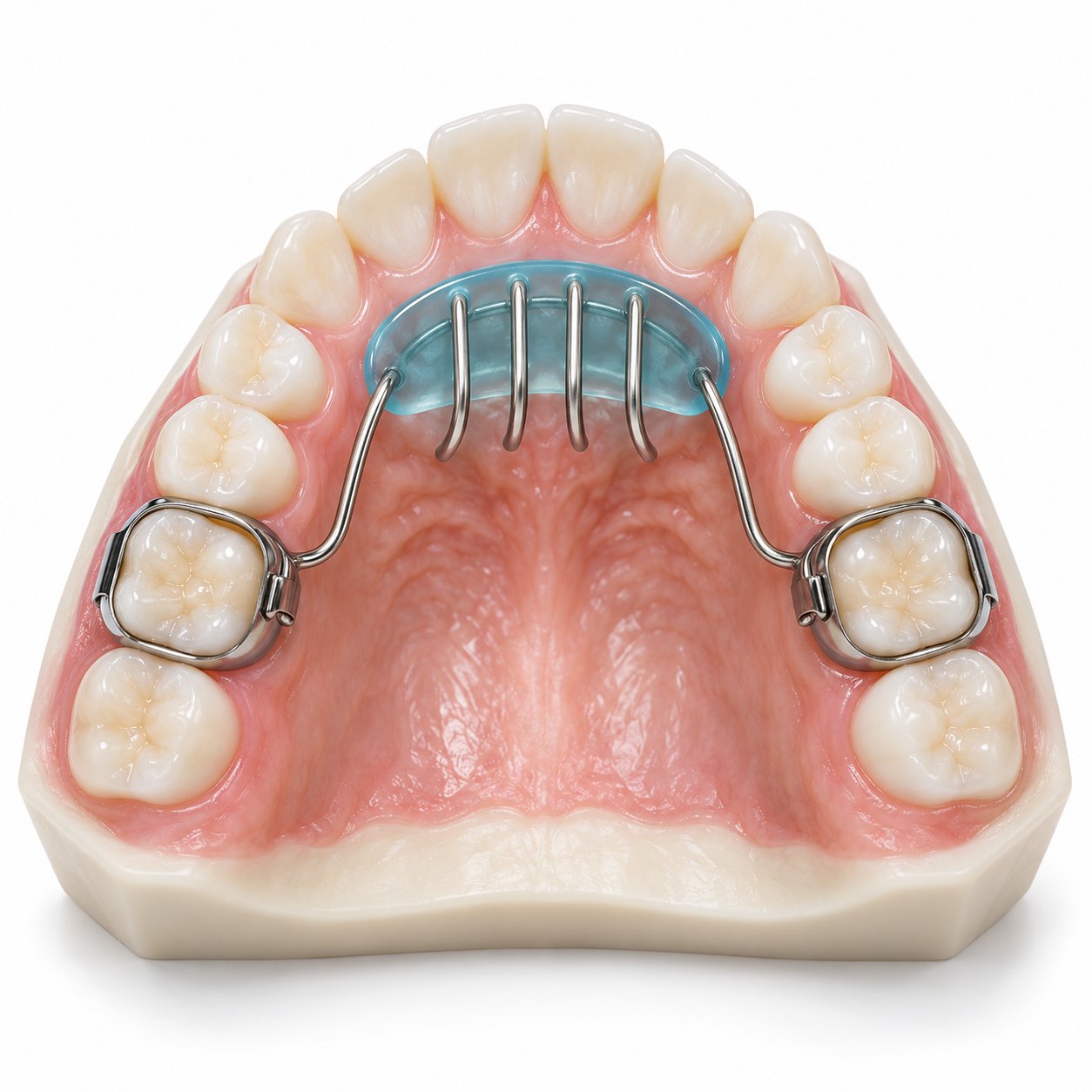
Pediatric Space Maintainers: These are fixed appliances such as band-and-loop or lingual arches which are placed immediately after premature loss of primary teeth to create space for the permanent tooth that has yet to erupt.
Surgical Extraction: This method is utilized if the buried tooth is so malformed, cystic, or close enough to other root tips that it creates a risk to surrounding healthy roots.
Prices of treatment in Kerala depend on how complicated your case is, the clinic you go to, and the type of material used:
Diagnostic X-Rays and CBCT Scan: ₹800 – ₹3,500
Pediatric Space Maintainer: ₹3,500 – ₹8,000
Laser Mucosal Exposure: ₹4,000 – ₹10,000
Surgical Exposure with Orthodontic Traction: ₹25,000 – ₹65,000 (includes appliance phase)
Cosmetic alignment of teeth is excluded, but part of the cost of surgery for serious impaction, cyst removal, trauma, etc., may be reimbursed through some private health insurance policies and public sector schemes in government dental colleges.
Pediatric Dentist (Pedodontist): The primary specialists for the early detection of issues that may arise during eruption and space preservation, as well as the management of eruption in children under age 7.
Orthodontist: Orthodontic treatment is necessary if traction, arch expansion, or comprehensive bracing are needed to position the teeth properly.
Oral and Maxillofacial Surgeon: This is the surgeon called in to remove cysts, manage deep bone impactions, and perform other complex surgical exposures.
Immediate (Within 24–48 Hours): There will be an immediate need to address this issue, as severe pain, facial swelling, fever, or visible displacement of adjacent teeth may be occurring because of the partially erupted tooth.
Prompt (Within 2–4 Weeks): It would be beneficial to have this addressed within four weeks if one permanent tooth has been asymptomatic in eruption for greater than six months versus its counterpart.
Elective / Routine: This should be monitored routinely during the regular bi-annual visits with your child’s pediatric dentist from age 6 through 12.
How long does it take for a permanent front tooth to come in once the baby tooth falls out?
Typically, it takes anywhere from 1 to 3 months for a permanent tooth to emerge when the baby tooth has fallen out. It is possible that if this happens over a time frame greater than six months, thick gum tissue, insufficient room for proper positioning, or other physical barriers such as an additional tooth germ could possibly be contributing to delayed emergence, at which point an X-ray will need to be taken to evaluate.
Are there specific types of candies or snacks available locally that would hinder tooth eruption?
Sticky, old-fashioned candy and similar traditional sweet treats do not prevent the process of tooth eruption, but contribute greatly to the development of extensive early childhood caries in baby teeth. Early caries destruction of baby teeth creates crowding of adjacent developing teeth, creating a physical barrier against incoming permanent teeth.
Would surgically exposing an impacted tooth be painful to the child?
Not at all. Pediatric dental care utilizes topical anesthesia gels to provide numbness, injectable local anesthetic to eliminate discomfort during surgery, and soft-tissue laser treatments. Any soreness experienced by the child post-surgery is usually minor and is generally manageable with pediatric analgesic syrup for 1–2 days.
Monitoring of eruptions between ages 6 and 8 is also important for determining if there are eruption delays. Low-dose digital X-rays taken during regular dental appointments will allow dentists to determine if there are any physical barriers that could delay an eruption. If a barrier exists, it can be easily removed with space maintenance and/or other laser-assisted treatments that may help avoid the need for complex jaw surgery in the future.



.jpg)
.jpg)
.jpg)
.jpg)

.jpg)
.jpg)
.jpg)
.jpg)
.jpg)
.jpg)
.jpg)
.jpg)
This site is part of the DEC Dental Network — a Dental Equity & Connectivity ecosystem of clinical, educational, and patient-support brands operated by Team Dental Pvt. Ltd. Our network works as one — guiding you from your first search to your final smile.
© 2026 Team Dental Pvt. Ltd. · Founded 2013, Angamaly, Kochi, Kerala
About DEC Dental