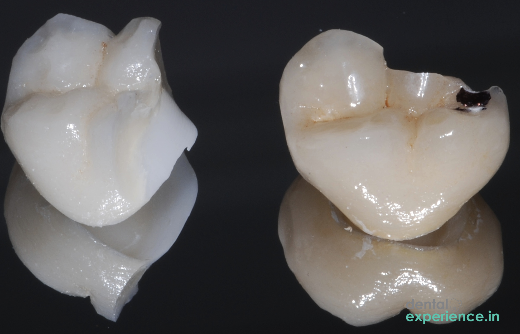
A ceramic crown fracture occurs when there is structural failure or breaking of a tooth-colored, biocompatible dental restoration. Ceramic crown fractures involve the clean break, chipping, or shearing of ceramic materials including lithium disilicate or zirconium dioxide which are bonded over a prepared natural tooth structure.
There are two classifications within the dental profession:
Uncomplicated Fractures: Only the ceramic material has chipped off, therefore the natural tooth structure or metal substructure remains intact.
Complicated Fractures: There is a break in the ceramic which has compromised the natural tooth structure or core build up material or exposed the delicate inner pulp chamber which contains the nerves and blood vessels.
When a crown fractures, it disrupts the uniform application of biting forces to your dental arch. As a result, your bite becomes unbalanced creating uneven wear on adjacent teeth, and exposes the margins (the precise junction where the edge of the crown meets your prepared natural tooth structure). When the margins become exposed, it allows for small spaces where bacteria can collect, increasing the potential for localized decay.
Clinical Definition: Physical breakdown, chipping, or total separation of a ceramic dental crown.
Indications that Require Urgent Care: Severe sharp pain upon inhalation of cold air, a loose or unstable crown, bleeding of the surrounding gums, swelling or exposed dark metal framework or nerve tissue.
Treatment Options: Polishing of intra-oral ceramic for small chips, repair via composite resin bonding or full replacement with premium, CAD/CAM milled monolithic zirconia crowns.
Potential Risks: Development of aggressive decay beneath the crown, irreversible nerve damage, and soft tissue lacerations from sharp ceramic edges.
Early detection of a ceramic crown fracture will reduce the chance of permanent nerve damage. Symptoms of a fractured ceramic crown vary but include both physical and sensory changes:
Sharp Edges: The patient experiences a new jagged texture or sharp edge that continues to irritate scrape cut etc. the sides of their tongue and/or interior of their cheek.
Localized Pain: The patient experiences dull aching while eating/chewing or a sharp electrical shock-like pain when biting down on hard objects.
Temperature Sensitivity: In vital (living) teeth, temperature changes trigger fluid movement within microscopic channels in the dentin (dentinal tubules), stimulating the nerve. Note that if the tooth previously underwent root canal therapy, it has no vital nerve tissue and will not experience temperature sensitivity.
Visible Changes: Cracks resemble spider webs on the surface of the tooth; parts of the crown are missing; a dark grey line appears along the gum line.
Misaligned bite: The patient consistently feels that their upper/lower teeth do not fit together correctly when their mouth is closed.
Although minor ceramic chips can often wait until your next scheduled appointment, there are several indicators that warrant an emergency visit to your dentist immediately:
Extreme Throbbing Pain: Nerve damage or severe pulp inflammation.
Swelling Along Gums: Active bacterial infection/abscess.
Crown Completely Loose: Immediate danger of ingestion/swallowing the free crown.
Additionally, if you notice an unusual odour/taste coming from around your fractured tooth, it is possible that bacteria are actively fermenting trapped food items under the free ceramic layer.
Fractures of a ceramic crown typically occur due to repeated mechanical stress or sudden trauma.
Trauma Caused by Biting on Hard Local Foods: Dental clinics in Kerala regularly treat crown fractures resulting from biting hard on unpopped corn kernels in Pori (puffed rice); stray stones found in traditional red rice varieties; and other hard fried Kaya Varuthathu (Banana Chips) and Sharkara Varathi.
Parafunctional Habits: Long-term chronic, unconscious nocturnal bruxism (teeth grinding) or day-time clenching causes extreme repetitive vertical force on the ceramic matrix leading to fatigue over time.
Fatigue of Porcelain Material: Micro-cracks form internally in early generation porcelain materials after many years of experiencing strong chewing forces.
Poor Cementation Techniques: Bubbles forming during placement of adhesive cements; uneven layers of adhesive cements; and lack of solid internal support provide weak points where the overlying ceramic collapses under normal chewing forces.
There are various risk factors that increase susceptibility to unpredictable failure of ceramic restorations:
Food Choices: Eating diets high in hard dense foods and/or constantly snacking on sticky sweet foods reduces strength in both the ceramic structure and its adhesive bond.
Heavy Teeth Grinding: Extreme, unrelenting pressure on your restorations caused by chronic teeth grinding.
Under-Preparation of the Tooth: Insufficient reduction of the original natural tooth structure during the shaping process leaves inadequate physical space for the ceramic, resulting in a restoration layer that is too thin and fragile to withstand normal chewing forces.
Anatomical Bite Issues: Maligned teeth direct abnormal uneven forces to a specific point on the ceramic crown.
Old Age of Restoration: Crowns constructed from older porcelain fused to metal (PFM) combinations are more susceptible to the ceramic layer shearing away from its metal base.
You can safely conduct this simple step-by-step self examination to verify your suspicion that your crown is cracked:
Step 1. Visual Examination: Use a clean mirror and flashlight.
Dry the area thoroughly with a clean cloth. Inspect closely for visible cracks; displaced portions; missing segments; or an exposed dark metal substructure.
Step 2. Tactile Evaluation: Carefully run your tongue along all surfaces (front/rear/bite) of the tooth. Document any areas that have sharp jagged edges or rougher than usual textures. Do not use your fingers or sharp objects to probe the tooth.
Step 3. Mobility Test: Using a clean cotton swab apply gentle pressure on each side of the crown. Determine if the crown shifts, moves, or tilts relative to the underlying prepared tooth structure. (Note: If the tooth has had a root canal, you may feel no pain even if the crown is completely loose).
Step 4. Sensitivity Documentation: Take sips of room temperature water slowly. Document whether sipping water elicits a dull ache; sharp flash of pain; or no response to discuss with your dentist
Your prosthodontist/GD uses systematic diagnostic tools to accurately assess extent of crown fracture:
Visual Evaluation: Using a fine-tipped explorer tool, the dentist maps out surface cracks & checks integrity of margins (boundary between crown & gumline).
Diagnostic Radiographs: Bitewings and intraoral periapicals are used to view root structure below the crown & detect hidden decay/hidden cracks going beyond gumline.
Transillumination: Exposing internal stress lines/fractures within the crown using transillumination technique. These cracks remain invisible when examined with Digital radiography alone.
Thermal Test: Assessing the vitality and sensibility of the tooth's internal nerve tissue (pulp) to determine if it is healthy, inflamed, or necrotic (dead).
Dental professionals categorize ceramic crown fractures into three distinct tiers of severity:
|
Severity Level |
Clinical Presentation |
Required Intervention |
|
Grade I (Superficial) |
Minor chipping limited strictly to the outer ceramic glaze; no metal or dentin exposed. |
Smooth contouring, intra-oral diamond polishing, or minor composite bonding. |
|
Grade II (Moderate) |
Extensive ceramic shearing that exposes the underlying metal coping or internal dentin matrix; causes mild temperature sensitivity. |
Complete removal and replacement of the crown, or complex composite resin bonding. |
|
Grade III (Severe) |
Catastrophic split extending beneath the gum line, accompanied by clear structural root fractures or direct exposure of the nerve pulp. |
Emergency crown removal and root canal therapy (if the root is structurally intact), or complete tooth extraction followed by a dental implant if a structural root fracture is present. |
The best option for restoring a damaged ceramic crown is by way of contemporary, high-quality restorative techniques:
Single piece Zirconia crowns are manufactured via the use of computer-aided design and manufacturing (CAD/CAM) technology. The crowns are made from a single solid piece of zirconium oxide. Due to its very high flexural strength of approximately 1200 MPa, it is highly resistant to chipping, though microcracks can still develop if it is adjusted improperly or subjected to extreme, unprotected grinding.
When aesthetics is the number one priority, lithium disilicate glass-ceramic is an unparalleled choice. It provides superior translucency and closely replicates the appearance of natural tooth enamel. Lithium disilicate crowns are chemically bonded to the tooth structure, providing a seamless aesthetic finish.
When a Grade III fracture occurs and damages the nerves within the tooth structure, an advanced laser-assisted endodontic procedure will be used to remove all bacteria from the root canals. This process is performed prior to constructing a new core build up and replacing the crown.
Dental treatment costs in Kerala differ depending upon the dental material chosen by each patient; whether or not your dentist has invested in state-of-the-art CAD/CAM technology and/or equipment to assist with treating dental emergencies; and how complicated the case may be:
CAD/CAM Monolithic Zirconia Crown Replacement: ₹ 9,000 - ₹ 18,000 per tooth.
Premium E.max Lithium Disilicate Crown Replacement: ₹ 11,000 - ₹ 22,000 per tooth.
Porcelain-Fused-to-Metal (PFM) Base Options: ₹ 3,500 - ₹ 7,500 per tooth.
Mild Cosmetic Repairs with Minor Composite Resin Bonding / Diamond Polishing: ₹ 1,500 - ₹ 3,500.
Selecting the proper dental specialist helps guarantee the successful outcome of your restoration:
Prosthodontists: Prosthodontists attend three additional years of advanced post-graduate university education and training focused solely on developing, fabricating, and properly fitting prosthetic teeth; bridges; and complex ceramic crowns. Prosthodontists are ideal experts to treat and replace fractured crowns.
Endodontists: When a Grade III fracture damages the nerve pulp or reaches deeply into the pulp chamber, an endodontist is needed to successfully complete root canal therapy to eliminate infection and preserve the tooth within the jaw.
General Dentists: General Dentists are qualified to evaluate and provide emergency care for your condition; smoothen out sharp edges on your ceramic crown; repair small cosmetic chips in your tooth using minor composite resin bonding; and arrange further treatment at a specialty clinic.
Your dentist will determine the length of time required to complete your crown restoration based upon the level of urgency associated with your particular situation:
Immediate (within two hours): Required for immediate treatment when there is bleeding; swelling; removal of a complete crown; or extreme, untreated pain associated with exposure of a nerve.
Urgent (within twenty-four to forty-eight hours): Essential for moderately severe Grade II fractures when internal dentin becomes sensitive to hot and cold temperature stimuli; or when sharp ceramic edges cut into your tongue.
Elective (five to seven days): Appropriate for extremely minor Grade I surface cracks resulting in mild cosmetic defects or slight texture irregularities.
Is it safe to fix my broken ceramic crown at home using common household adhesives or over-the-counter superglue?
Never attempt to attach a cracked ceramic crown back onto your tooth using common household adhesives or over-the-counter superglue products. Household adhesives or over-the-counter superglue products contain toxic chemical compounds that can irreparably damage the healthy pulp inside your living tooth. Additionally, both types of adhesive create an uneven layer that distorts your natural bite alignment, ultimately cracking the remaining healthy roots of your natural tooth.
What is the safest amount of time I can wait before seeing my dentist about my broken crown?
You can wait five to seven days without concern for a scheduled appointment if you do not have any symptoms including pain, swelling, or sensitivity. If your exposed dentin layer is open to bacteria colonization, however, delaying an appointment for more than just a couple of days could convert what would have been a simple crown replacement into a complex and costly root canal treatment.
Will a monolithic zirconia crown look as good as my existing front teeth?
Yes. Although earlier versions of zirconia had flat and chalky appearances, current multi-layered monolithic zirconia combines outstanding mechanical properties with realistic translucence. Your laboratory technician can accurately replicate the gradient color changes found in your surrounding natural teeth to create an incredibly natural appearance regardless of light conditions.
When managing a ceramic crown fracture, our primary goal is to protect the underlying natural tooth structure and restore a balanced bite. Modern dental technology has shifted from older porcelain-fused-to-metal crowns toward exceptionally strong materials like monolithic zirconia and lithium disilicate.
Long-term success depends on an accurate diagnosis using digital radiography and transillumination lights. Patients must avoid fixing broken crowns at home and seek immediate professional care to prevent deep bacterial contamination. Consistent oral hygiene, routine dental checkups, and wearing custom night guards if you grind your teeth are vital to ensuring your premium restorations last for decades.














.jpg)
.jpg)

.jpg)
.jpg)

.jpg)
.jpg)
.jpg)
.jpg)
.jpg)
.jpg)
.jpg)
.jpg)