

Fractured Baby Tooth (Primary Tooth Fracture) is caused by physical trauma to the tooth, resulting in loss of structure. Fractured primary teeth range from minor enamel chip fractures to major crown-root fractures where the succedaneous (permanent) tooth germ may be compromised.
Primary teeth consist of an enamel layer on the outside; a dentin layer underneath the enamel; and at the center is a pulp chamber. The pulp chamber contains vital nerve endings and blood vessels. The enamel layer is much thinner, and the pulp chambers are much larger in primary teeth compared to permanent teeth. This makes a small crack in the enamel layer a large risk factor for infection, as bacteria can easily penetrate into the pulp through the crack.
Clinical Definition: Direct or indirect force causing a fracture of a primary (baby) tooth.
Indicators of Urgency: Bleeding from within the tooth, severe pain/throbbing, exposed pink/red tissue within the fracture, mobile fragments of tooth, and abscess formation/gum swelling.
Recovery Options: Smoothing sharp edges (smoothing); composite resin bonding; pulpotomy (baby root canal); stainless steel/pediatric zirconia crown; or extraction with space maintainer.
Children who have suffered a fractured tooth may exhibit visual, tactile, and behavioral signs that parents/caregivers will see when their child is fed/brushed:
Visual Signs: A fractured tooth will have jagged edges visible on the tooth crown as a result of enamel loss.
Color Change: The color change will be an indicator of what is happening to the pulp. If it changes to a light pink color, then there has been acute pulp hemorrhaging. If the color becomes darker (gray/black), then there has been necrotizing pulpitis occurring.
Sensitivity to Temperature & Movement: Because of exposed dentinal tubules, the child will cry and/or refuse to take cold liquids or drinks (payasam), hot food or beverages, and/or sweet items.
Aversion to Mastication: Due to masticatory pain, the child will avoid chewing on one side of his/her mouth. In addition, he/she will drop all firm foods such as tapioca (kappa) and hard banana chips (nendran chips).
Tissue Changes: Due to sharp fractured tooth fragments, the child can experience lacerations to the inner lip/tongue/buccal mucosa.
Active Bleeding from Fractured Pulp: Blood will ooze from the fractured area of the tooth.
Swollen Gingiva / Sinus Tract(s): Smaller than normal bumps/gum pimples (parulis) are forming near the root of the fractured tooth and draining pus.
Mobility of the Teeth: The fractured teeth will move back and forth horizontally and vertically more than they should.
Fracture or Displacement: The fractured tooth can become displaced downward (intrusive luxation) or sideways (lateral luxation) from its original position.
General Health Symptoms: Fever, facial asymmetry, etc.
Childhood traumatic injuries are very prevalent due to physical damage that can occur in children as they develop physically:
Physical Injury from Unintentional Falls: Children developing their gross motor skills fall off wet tiles at home, while walking up verandah stairs, or other similar accidents that could cause them to hit their face against objects.
Injury from Recreational Activities: Riding bicycles, playing on swing sets, or participating in team sports where there is no protection for the mouth, such as wearing a mouthguard.
Biting Down on Hard Foods: Biting into hard foods or hard items—i.e., food debris such as seed pits, candies, or certain local snack foods (such as dry achappams or murukkus).
Impact by Foreign Objects: Biting down with an excessive amount of force into plastic toys, metal feeding utensils, or glass containers.
A variety of structural and behavioral factors exist which will increase the likelihood of a child experiencing a fracture in one of their primary teeth:
Class II Malocclusion (Protruding Upper Front Teeth): Also referred to as "buck" teeth; these protrude beyond the upper lip, providing little to no protection when the child falls onto their front teeth. The force that hits the buck teeth when the child falls can be up to 80% greater than if their buck teeth were covered completely by the upper lip.
Deep Decay on a Tooth (Early Childhood Caries): Decay reduces the structural integrity of the enamel on the outside of the tooth, which makes it susceptible to cracking under normal masticatory forces.
Hypoplastic Enamel: This condition occurs when the enamel forming on the outer layer of the tooth is inadequate. The result is a thinning of the enamel, which creates a weakened structure. When subjected to normal chewing pressures, this thin enamel structure will eventually crack.
Increased Hyperactivity: An increased risk of injury from uncoordinated play increases the likelihood of a child experiencing dental trauma.
When you have the opportunity to evaluate the amount of damage that has occurred to your children’s teeth at home and do not want to make things worse:
Clean Your Hands: Before even going near the inside of their mouths, wash your hands with soap and water.
Stop the Bleeding: Have them bite on a clean wet gauze pad or roll for about five minutes. Gently pull back their upper or lower lip under bright light. Look at the location where the tooth was broken. Identify the layers exposed by the break:
Only White: Only the enamel fractured.
The Yellowish Middle Layer Can Be Seen: Dentin fractured.
A Red Spot in the Center: Pulpal area is exposed; requires immediate emergency treatment.
Check How Stable the Fragments Are: Gently place your clean finger over the tooth to determine whether it will stay put or come out.
Warning: Do not push or pull on a loose piece.
Pediatric dentists use specific diagnostic methods to assess the extent of the damage:
Visual & Tactile Examination: Visual and tactile examination of the fracture line(s), sharp edge(s), and soft tissue laceration(s) via a dental mirror and explorer.
Intraoral Radiographs: Periapical X-rays to assess root health, alveolar bone involvement, and proximity of fracture to developing succedaneous (permanent) tooth germs.
Transillumination: A high-intensity light beam passed through the tooth to reveal hidden craze lines (microfractures).
Palpation & Mobility Tests: Lightly apply pressure to determine if there is inflammation or disruption of the periodontal ligament.
|
Severity Level |
Structural Damage |
Primary Clinical Sign |
Immediate Action |
|
Grade I (Craze Lines) |
Micro-cracks in enamel only |
Visible thin lines, no lost tooth structure |
Elective monitoring |
|
Grade II (Uncomplicated Crown FX) |
Enamel and dentin involved |
Yellowish dentin visible, cold sensitivity |
Restoration within 24–48 hours |
|
Grade III (Complicated Crown FX) |
Enamel, dentin, and pulp exposed |
Red dot in center, active pain/bleeding |
Emergency care within 2–4 hours |
|
Grade IV (Crown-Root Fracture) |
Involves enamel, dentin, and cementum extending below the gum line (with or without pulp exposure) |
Fragment loose, gums bleeding profusely |
Immediate specialist evaluation |
Options in pediatric restorative dentistry now include the ability to save functional form while also preserving the potential of the permanent successor:
Fluoride Varnish & Enamel Smoothing: Mild enamel chips will be addressed by smoothing rough edges and applying a high-concentration fluoride varnish to reduce sensitivity.
Composite Resin Bonding: Nano-hybrid composite resin bonded to restore tooth shape in one visit using tooth-colored restoration.
Pulpotomy: If the pulp is exposed, remove the infected portion of the upper pulp tissue, treat the remaining healthy pulp tissue with therapeutic medications such as mineral trioxide aggregate, and seal it.
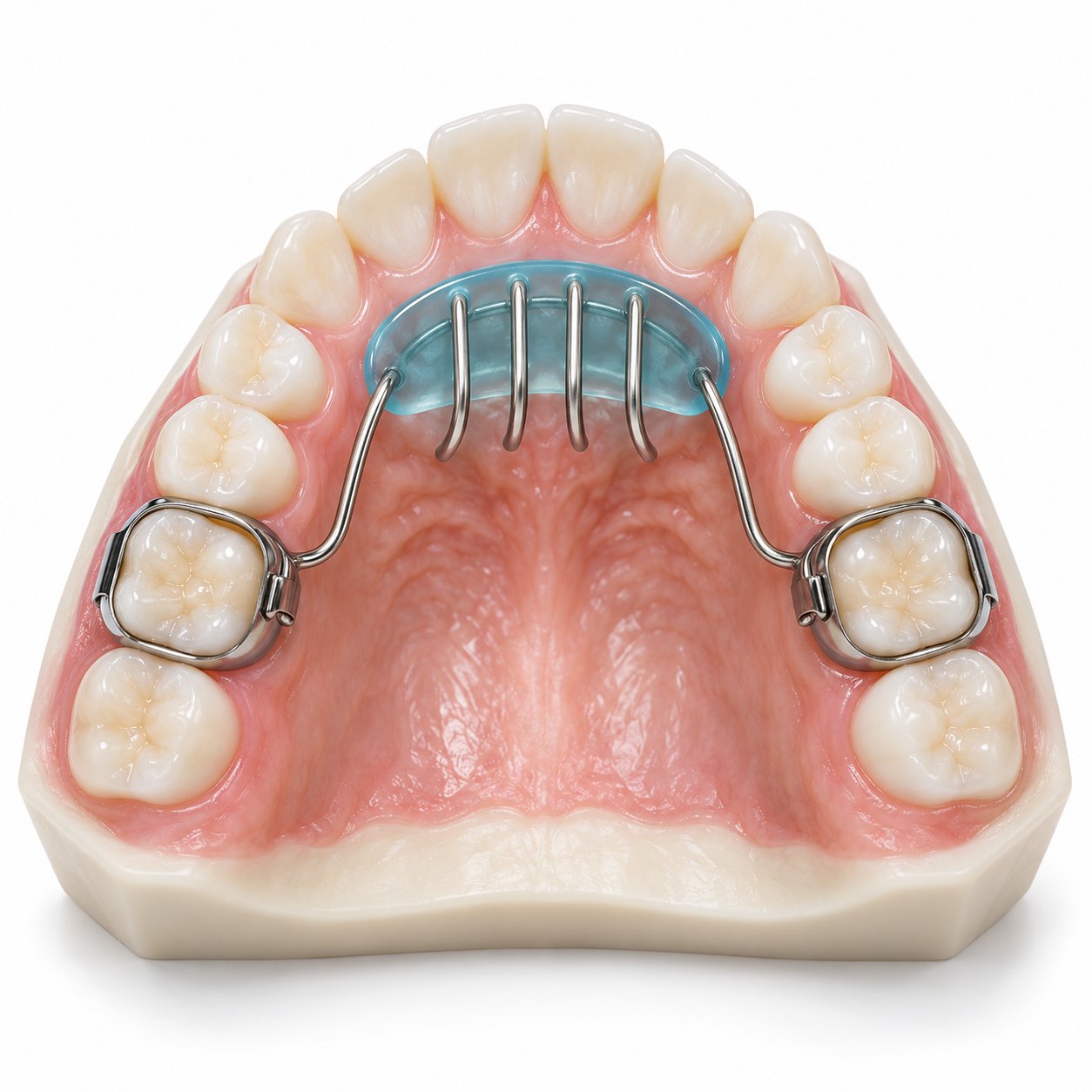
Pediatric Crowns: Biocompatible preformed crowns made from stainless steel for molar teeth or full-coverage zirconia crowns for front teeth will be placed on severely fractured baby molars or front teeth.
Extraction: In cases where the fracture extends too far down into the root or there has been extensive infection, extract the tooth to protect the developing permanent tooth germ.
Dental treatment cost varies based on the area you live in (urban dental offices located within Kochi vs. rural dental office locations) as well as the type of materials used for your treatment and the level of experience of your dentist.
Consultation/X-Ray: ₹400–₹1,200
Enamel Smoothing/Fluoride Application: ₹800–₹1,500
Composite Resin Restoration: ₹1,500–₹3,500 per tooth
Pediatric Pulpotomy: ₹3,000–₹6,000
Zirconia/Aesthetic Primary Crown: ₹5,000–₹9,500
Extraction/Space Maintainer: ₹2,500–₹6,000
The majority of people pay out of pocket; however, major corporate medical insurance companies and regional empaneled programs continue to provide financial support for emergency pediatric trauma situations.
General Dental Practitioner: Suitable for primary triage, simple smoothing, and minor enamel composite fillings.
Pediatric Dentist (Pedodontist): Recommended for primary tooth fractures. They are specialists in child behavior management, pulpal therapies, space maintenance, and growth-oriented care.
Pediatric Endodontist / Endodontist: Needed when there is complex pulpal revascularization or root-level fracture.
Oral & Maxillofacial Surgeon: Needed when there is severe facial trauma along with the dental fracture that involves jaw fractures or complex soft-tissue suturing.
Emergency Window (2 to 4 Hours): Recommended for Grade III complicated fractures with exposed pulp or displaced teeth. Quick coverage will prevent irreversible pulp necrosis and deep bacterial contamination.
Urgent Window (24 to 48 Hours): Used for Grade II dentin fractures. Prompt coverage will protect exposed dentinal tubules from bacterial invasion and stop thermal pain.
Elective Window (Within a Week): Can be addressed during routine clinic hours; superficial enamel cracks or minor roughness without pain.
Should my child's blackened baby tooth be removed right away because it was damaged for a little over a week?
No. The most likely cause of a darkening of a fractured baby tooth is internal hemorrhage or pulp death (similar to a bruise), and the dentist will use an X-ray to evaluate whether there has been a loss of bone structure as well as infection in the surrounding tissue. If there are no signs of active inflammation or loosening of the tooth, the tooth can be observed instead of being removed.
If you do nothing about your child’s seemingly painless chipped baby tooth, what will happen?
Even though some chips in baby teeth don’t produce any symptoms, they can create pathways for bacteria to enter through tiny tubes called dentinal tubules into the pulp, creating latent infection, possible abscesses, along with possible scarring or discoloration of the developing permanent tooth below.
Will glue hold together a fragment of a broken baby tooth like an adult tooth?
Although it is common to reattach fragments of permanent teeth using composite resin, reattaching fragments of baby teeth is almost never successful due to a very thin layer of enamel and a relatively small surface area. Restoring broken primary teeth with a composite resin restoration or aesthetic pediatric crowns provides a much longer-lasting repair.
Trauma to pediatric dentition requires prompt clinical assessment regardless of the degree of apparent damage to the untrained observer. As primary teeth provide critical guidance for permanent teeth to develop properly, maintain the normal structure of the jaw, and contribute to both adequate nutrition and proper speech patterns, neglect of a fractured primary tooth will significantly increase the risk of bacteria infecting the permanent tooth bud that develops in direct contact with the roots of the fractured primary tooth. Following any type of traumatic incident, parents should obtain an official clinical examination and radiograph within 24–48 hours to help promote ideal oral health results over time.





.jpg)
.jpg)
.jpg)
.jpg)
.jpg)
.jpg)
.jpg)
.jpg)
.jpg)
.jpg)
.jpg)
.jpg)
This site is part of the DEC Dental Network — a Dental Equity & Connectivity ecosystem of clinical, educational, and patient-support brands operated by Team Dental Pvt. Ltd. Our network works as one — guiding you from your first search to your final smile.
© 2026 Team Dental Pvt. Ltd. · Founded 2013, Angamaly, Kochi, Kerala
About DEC Dental