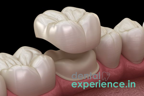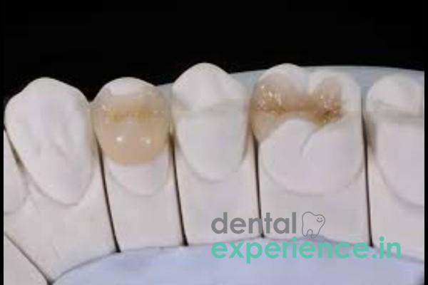
A fractured inlay represents a structural failure of a customized, indirectly fabricated dental restoration that is placed within the internal grooves of a tooth, specifically between the cusps rather than covering them, to repair structural damage. An inlay differs from other types of composite fillings because it is made in one complete piece. Customized inlays can be made using a variety of materials including, but not limited to, gold, porcelain and/or composite resins. The inlay restoration is then bonded to the tooth's internal dentin structure and peripheral enamel margins.
When an inlay fails structurally, the overall integrity of the tooth is compromised. Dental failures of this nature fall into the category of "advanced" restorative dental failures. When an inlay fails, the protective dentin layer is left exposed to the oral cavity, which can eventually lead to pulp chamber involvement if left untreated. As a result, the exposed dentin and underlying structures are subject to invasion by oral bacteria, thermal shock, and mechanical force. Therefore, prompt clinical action should be taken to protect against tooth loss.
Clinical Definition - Structural failure of a custom, indirect dental restoration positioned within the internal grooves (between the cusps) of a tooth.
Indications of Emergency Action - Sudden onset of localized pain while eating hard/rough textured foods, continuous throbbing pain, temperature sensitivity to warm/cold liquids/drinks, visible crack(s) or loose restoration fragment(s).
Treatment Options - Composite bonding to repair minor chips, replacement using CAD/CAM ceramic technology, conversion to an onlay or full-coverage crown, and endodontic therapy if the pulp was breached.
Risk Considerations - Development of secondary caries in the affected tooth, deep infection of the pulp chamber, and possible vertical fracture of the tooth.
Common symptoms of patients experiencing a fractured inlay include:
Pain: Patient reports acute onset pain when chewing firm/ rough textured food.
Sensitivity to Temperature: Persistent heat/cold ache associated with drinking fluids at room/warm temperatures.
Textural Abnormalities: The tongue identifies sharp edges or abnormal gaps on the occlusal or interproximal surfaces of the molar.
Mobility of Restoration: Sensation of movement of the inlay during speech/mastication.
Minor chip repairs can often wait for a scheduled appointment. However, there are several signs that indicate urgent/emergency care may be needed immediately. These include:
Continuous/Burning Pain: Fracture propagation directly into or extremely close to the pulp chamber has allowed bacterial invasion, resulting in irreversible pulpitis.
Abscess/Fistula: A small swelling or painful bump on the gum adjacent to the affected tooth indicates localized spread of a bacterial infection from the pulp into the surrounding bone and soft tissue.
Active Bleeding: Bleeding occurs primarily from the surrounding gingival tissue due to trauma from sharp restoration fragments or food impaction.
Foul Odor/Taste: Ongoing presence of bad taste/suggests that food debris and bacteria continue to decompose under the loose/debonded restoration.
Traumatic Mastication: Chewing extremely difficult local foods i.e., uncooked hard banana chips fried in coconut oil or biting down on unpopped kernels/kernels in unprocessed rice.
Fatigue of Material: Repetitive chewing motions produce microscopic fractures in both older composite resins and porcelain restorations eventually producing catastrophic failure.
Secondary Decay: Bacterial leakage under the margins of the inlay leads to continued demineralization of the tooth structure supporting the inlay until the tooth base collapses.
Parafunctional Habits: Nocturnal bruxism/daytime clenching habits place excessive vertical/lateral forces on the restoration.
Initial Tooth Preparation Size: Patients who have lost most of their original tooth structure/enamel are much more likely to develop flexure-induced stress on their inlay bonding agent.
Inlay Material Type: Early-generation composite resins and traditional dental ceramics were inherently more brittle than modern high-strength materials like lithium disilicate (E.max) glass-ceramics and solid zirconia.
Eating Habits: Sticky/chewy Indian sweets such as halwa or frequent consumption of hard snack products accelerate adhesive deterioration.
Orthodontic/Dental Relationship: Poor occlusal alignment or malalignment that applies additional excessive load directly at the restoration-tooth interface.
If you think your restoration has failed, you can perform the following safe, step-by-step visual and tactile self-assessment in front of a well-lit mirror:
Visual Inspection: Clean and dry the area gently with a clean tissue. Check for visible cracks, dark lines around the borders of the filling, or missing segments of the restoration.
Tongue Sweep: Gently run the tip of your tongue over the chewing surfaces of your molars to feel for sharp ridges, sudden dips, or loose pieces.
Pressure Test: Using a clean cotton swab, apply light, downward pressure to the center of the suspected tooth. Note if there is any movement or pain upon release.
Do not attempt to scratch the area with toothpicks, pins, or metal tools, as this can worsen the fracture or push bacteria deeper into the pulp chamber.
A definitive diagnosis requires a clinical evaluation by a dentist utilizing specific tools:
Visual and Tactile Examination: The dentist uses a specialized dental explorer to check the marginal integrity of the inlay and detect micro-leakage.
Digital Radiography: Intraoral X-rays identify secondary decay beneath the inlay margins and evaluate surrounding bone health, though standard 2D films have limited visibility for detecting early, un-displaced root fractures.
Transillumination: High-intensity light is passed through the tooth structure to illuminate hidden fracture lines that do not show up on standard X-rays.
Pulp Vitality Testing: Thermal or electrical stimuli are applied to confirm whether the nerve tissue inside the pulp chamber remains healthy and viable.
|
Severity Level |
Structural Damage |
Required Clinical Action |
|
Grade 1: Superficial |
Minor chipping of the inlay margins without loss of adaptation or dentin exposure. |
Conservative polishing or micro-hybrid/nano-hybrid composite resin smoothing. |
|
Grade 2: Moderate |
Structural fracture of the inlay body; dentin is exposed but the pulp chamber is secure. |
Complete removal of the failed inlay and replacement with a new CAD/CAM restoration. |
|
Grade 3: Severe |
Extensive fracture of the restoration that propagates into the natural tooth structure, fracturing the structural cusps and exposing or approximating the pulp chamber. |
Root canal therapy followed by full structural coverage with a porcelain or zirconia crown. |
There are several ways to restore a broken inlay:
New CAD/CAM Ceramic Onlays/Inlays - The latest advancements in dentistry have made it possible to create a brand-new inlay using intra-oral scanning and milling from a block of lithium-disilicate ceramic. Lithium-disilicate ceramic closely mimics the aesthetic translucency, wear resistance, and physical properties of natural tooth enamel and can be fabricated and placed within a single visit.
Full Coverage Zirconia Crown - Due to excessive structural failure of the tooth, the tooth will need to be shaped to accommodate a full zirconia crown providing 360 degree protection for chewing forces.
Direct Biomimicry Nano-Hybrid Resin - Minor fractures along the margin can be sealed using special nano-hybrid composites directly applied to the defective area without removing the original inlay.
The cost of restoring a broken inlay will vary based upon the type of material used and how damaged the tooth is. With modern CAD/CAM ceramics such as E-Max, most clinics charge between ₹8000 – ₹18000 per tooth for a replacement.
If a patient needs a full coverage zirconia crown due to excessive loss of tooth structure, the price usually ranges from ₹10000 – ₹25000. Additionally, if a root canal treatment is required before restoration, this additional cost may be anywhere from ₹4000 – ₹9000.
Typically, standard dental insurance programs classify inlays as major restorative procedures rather than basic fillings; consequently, plans may only offer partial coverage or apply an alternative benefit clause, requiring patients to pay the remaining balance out of pocket.
A General Dentist is fully qualified to diagnose a fractured inlay, place sedative temporary restorations, perform root canals, design CAD/CAM ceramic replacements, and place full-coverage crowns.
A prosthodontist is a dental specialist focused on the restoration of oral aesthetics and function, routinely managing advanced CAD/CAM ceramic replacements, complex onlays, or full-mouth bite rehabilitation.
An Endodontist is a root canal specialist and his/her services are recommended whenever the fracture compromises the pulp chamber resulting in significant nerve irritation or periapical infections.
Immediate Care Needed Within 2-4 Hours: Severe continuous pain, visible gum swelling, and/or a completely dislodged inlay leaving a deep, unprotected cavity in the tooth with associated bleeding from the irritated surrounding gum tissue.
Care Needed Within 24-48 Hours: Broken piece of inlay resulting in mild-moderate sensitivity, but no persistent throbbing or acute swelling.
Routine Follow-up Appointments Within 7 Days: Cosmetic chipping of the edges of the inlay that causes no pain and no thermal sensitivity, but results in an unappealing appearance to the tongue.
What should I do if my dental inlay breaks while eating?
Gently rinse your mouth with warm salt water to remove all debris. Store any pieces of broken inlay you were able to retrieve safely and avoid chewing on that part of your mouth until you can get evaluated by a dentist. Do NOT try to glue or push the piece back into place with your fingers.
How much will it cost me to repair a broken ceramic inlay?
Using a premium CAD/CAM ceramic restoration to replace a fractured inlay typically ranges between ₹8,000 and ₹18,000 based upon the type of equipment your dentist uses and the amount of tooth structure lost.
Can a fractured inlay be repaired with just a small composite filling?
Yes. If the break is just a minor chip along the outside edge of the inlay, your dentist can likely use some form of direct composite resin to “patch” over the defective area. However, if the main body of the inlay has actually split apart, you will require a complete new restoration of that tooth to stabilize it structurally.
Fractures represent far more than simply an aesthetic issue — they indicate an inherent compromise of a tooth’s structural mechanics. When either adhesives fail or material matrices fail, dentin loses its protective barrier against normal acidic processes occurring daily in our mouths as well as heavy mechanical load-bearing functions.
To achieve long-term success in preventing these failures relies heavily on proper selection of materials combined with regular preventative maintenance. To evaluate potential micro-leaks around restoration margins, patients should have routine examinations performed at least every six months. It is also important for patients to modify their diet (e.g., eat less frequently foods that tend to be hard). Also, utilizing protective night guards during sleep is critical to extending the functional life-span of more advanced dental restorations.














.jpg)
.jpg)

.jpg)
.jpg)

.jpg)
.jpg)
.jpg)
.jpg)
.jpg)
.jpg)
.jpg)
.jpg)