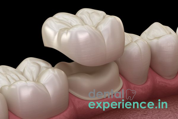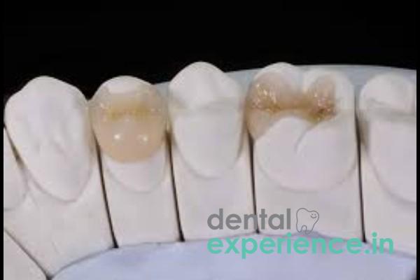
A fractured onlay represents a structural failure of a custom-made partial dental restoration created to restore the chewing surface of a tooth, differing from a full-coverage crown. An onlay is different from a filling which rests internally within the tooth structure, and a crown which fully encases the entire tooth. An onlay partially covers one or multiple cusps (raised areas on the chewing surface) of the tooth. Upon fracturing of the onlay, the structural integrity of both the enamel (the outer protective layer) and the dentin (the primary underlying mineralized structure protecting the nerve) is compromised. This is considered a type of complex restorative failure in clinical dentistry. Due to the nature of the fracture, the tooth will be highly susceptible to microleakage (bacteria seeping past the remaining restoration) potentially compromising the internal pulp chamber (nerve and blood supply) of the tooth.
Clinical Definition: Structural failure resulting in partial or total loss of function of a pre-fabricated porcelain, composite or gold partial crown restoration.
Urgent Indications: Acute severe pain while chewing; sudden onset of increased sensitivity to temperature extremes (hot/cold); visually apparent cracks; or removal of all or part of the ceramic portion.
Recovery Options: For minor surface chipping, direct composite bonding may be utilized; for deeper structural fractures, replacement with modern lithium disilicate (e.max.) onlays; or full coverage zirconia crowns may be necessary for significant fractures extending into the root.
Common symptoms patients experience due to a fractured onlay include distinct sensory and physical experiences:
Sensitivity: A sharp piercing sensation experienced immediately upon contact with hot beverages, cold liquids, or sweet items such as traditional payasam.
Pain: A transient sharp pain that occurs when biting down, or characteristically a sharp pinch experienced upon the sudden release of chewing pressure (indicative of a fracture line propagating into the dentin).
Roughness: A jagged and rough surface that is detected by the tongue and causes continuous irritation or micro cuts to the cheeks.
Visual Symptoms: Dark staining lines across the chewing surface of the tooth; visible absence of ceramic; noticeable difference in occlusion (contact) between upper and lower teeth.
Although a small chip in a porcelain restoration is an elective procedure, certain clinical indicators warrant urgent treatment. These indicators are indicative of a potential need for immediate emergency care:
Persistent Spontaneous Throbbing Pain: Persistent throbbing pain that prevents sleeping indicates the fracture has progressed far enough to result in irreversible pulpitis (irreversible nerve damage).
Gum Swelling: Development of swelling adjacent to the affected tooth at the gum line, which may indicate either an infectious process or an active abscess formation.
Mobility: Mobility or movement of the remaining natural tooth structures upon gentle touch.
Unpleasant Taste/Odor: Prolonged unpleasant metallic or putrid tastes, which suggest significant microleakage and/or active decay occurring under the displaced restoration.
Fracture of an onlay is rare and always caused by some form of mechanical stress combined with long-term wear and tear (fatigue):
Traumatic Bites: Unexpectedly encountering hard objects during eating - examples include accidentally biting down on an un-husked grain hidden in cooked rice, or biting down on a hardened piece of dried banana chips.
Fatigue Failure: Gradual expansion of microscopic structural weaknesses within older composite or porcelain restorations due to prolonged high frequency chewing ultimately results in catastrophic failure.
Microbial Degradation/Decay: Formation of new cavities along the marginal boundaries (periphery) of existing restorations, thereby undermining their integrity and contributing to structural collapse under normal chewing loads.
There are several predisposing risk factors associated with an increased likelihood of an onlay breaking:
Bruxism: Unconscious grinding/clenching behaviors during sleep contribute significantly to excessive forces applied against restorations.
Food/Beverages: Consuming large amounts of fibrous food sources (raw tapioca, etc.), sticky candies/sweets or tough meat products subject teeth to extreme lateral forces.
Initial Large Cavities: Teeth requiring larger initial restorations due to limited residual enamel are structurally weaker and therefore more likely to fail due to structural bending/flexural deformity and subsequent secondary cracking.
If you believe your onlay has broken, you may safely assess yourself with these procedures in front of a well lit mirror:
Visual Inspection: Use gauze to dry the area thoroughly then inspect visually for absent segments of ceramic, dark staining around the periphery, or an evident gap between tooth and onlay surfaces.
Tongue Sweep: Gently run the tip of your tongue along the chewing surface to detect irregularities or sharp edges/large craters.
Pressure Assessment Using Cotton Swab: Apply gentle downward pressure on separate portions of the tooth using a cotton swab. Identify areas where you feel a sharp pinch/pain when releasing pressure.
Warning: Do not use any instruments/metallic devices (toothpicks, pins, etc.) to scrape away fragments from the area. Avoid attempting to remove loose ceramics since this may further compromise the health of the underlying tooth structure.
The diagnosis of an onlay fracture is confirmed through an examination performed by a trained clinician utilizing precise diagnostic methods:
Visual/Tactile Examination: The clinician uses an explorer to verify the structural integrity of the margins and to detect any catching points or small cracks in/on the surface.
Diagnostic Imaging (Radiographic): Digital intraoral radiographs will help determine if there exists any evidence of secondary caries beneath the restoration or if the fracture extends into the root system.
Pulp Vitality Testing: Utilize thermal or electrical stimulation testing to assess the health status of the dental pulp, which dictates whether a straightforward replacement of the restoration is viable or if endodontic root canal therapy is required.
Dentists classify fractured onlays into three primary tiers:
|
Severity Level |
Structural Damage |
Primary Clinical Action |
|
Grade 1: Superficial |
Minor chipping confined strictly to the porcelain layer; no natural tooth tissue involved. |
Smoothing down rough edges or minor composite polishing. |
|
Grade 2: Moderate |
Structural break involves both the onlay and natural enamel/dentin, but the pulp chamber is unexposed. |
Complete removal of the failed restoration and replacement with a new onlay. |
|
Grade 3: Severe |
Deep fracture extending beneath the gum line or exposing the pulp chamber. |
Endodontic therapy (Root Canal) followed by a full-coverage protective crown. |
On a purely clinical basis, fractured onlays should never be considered as merely material failures. Rather they indicate potential underlying biomechanical stress or structural compromise. The earlier a fractured onlay is identified, the better the prognosis. Early detection allows preservation of important tooth structures and avoidance of costly endodontic therapy when required.
Success over the long term ultimately rests upon patient cooperation specifically through modification of diet habits in order to cease biting unyielding items and adherence to strict, twice daily oral hygiene regimens combined with semi-annual clinical assessments.














.jpg)
.jpg)

.jpg)
.jpg)

.jpg)
.jpg)
.jpg)
.jpg)
.jpg)
.jpg)
.jpg)
.jpg)