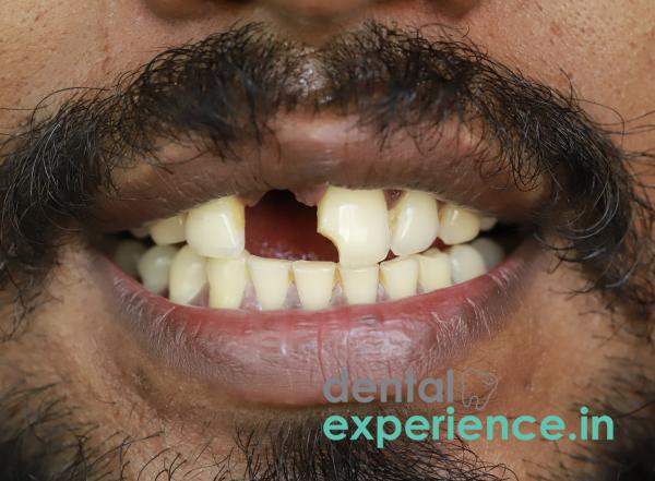
An avulsed tooth, defined as a tooth completely displaced from the alveolar socket and therefore losing all connection to the alveolar bone and periodontal ligaments, is among the most serious types of traumatic injuries to the teeth. The tooth is no longer anchored to the surrounding bone by means of the periodontal ligament (a collection of collagen fibers); in addition, the neurovascular bundle supplying the internal pulp chamber is totally severed. As part of a broader definition of dental emergencies, an avulsion is considered an urgent maxillofacial problem that requires immediate replantation in order to preserve the natural tooth structure.
Clinical Definition: Total displacement of a tooth from its alveolar socket, resulting in loss of support to both the bone and the nerve connections to the tooth.
Warning Signs: Heavy, non-stop bleeding from the socket; intense jawbone pain; and noticeable cracks, chips, or looseness in the surrounding teeth.
Treatment: The primary goal is to put the tooth back in the socket immediately and secure it with a flexible splint within the first hour. If the tooth cannot be saved, long-term options include a dental implant, a bridge, or a temporary partial denture.
A patient who has experienced an avulsed tooth will exhibit several distinct visual, tactile, and sensory symptoms associated with the injury:
Empty Alveolar Socket: There is a dark space where the tooth used to be located in the dental arch and there is active bleeding from the sockets of the tooth.
Continuous Localized Hemorrhage: Ongoing bleeding from the torn capillaries of the gingiva (gums) and/or the deeper walls of the socket.
Severe Pain: Unbearable pain in the jaw area caused by severed nerve endings at the base of the empty socket.
Edema of Soft Tissues: Rapid onset of inflammation and swelling of the surrounding areas including gums, lips, and face due to inflammatory response in the affected area.
Mobility of Adjacent Teeth: Loose or sensitive teeth adjacent to the injured tooth caused by transmission of impact energy throughout the dental arch.
Although missing a tooth is very disturbing, certain clinical signs warrant immediate consultation by an oral surgeon or emergency medical services:
Continued Uncontrolled Bleeding From Socket: Bleeding that cannot be controlled after application of steady pressure with sterile gauze for over 30 minutes.
Fracture of Surrounding Jawbone: Visible deformities or shifting of the jawbone and/or misalignment of portions of jawbone upon gentle touch to the affected area.
Loss Of Sensory Function: Numbness, tingling or loss of sensation extending to include parts of the lower lip, chin or cheek suggesting severe damage to deep nerves.
General Systemic Disturbances: Dizziness, vomiting, confusion and/or increased temperature may indicate possible concussive events or systemic infection.
Avulsions occur primarily as a result of mechanical forces acting on either directly or indirectly on the maxillofacial area:
Motor Vehicle Collisions: High speed collisions while riding two wheeled vehicles or falling from these vehicles onto highways. Often riders or passengers wear no helmet protecting the entire head.
Traumatic Contact During Competitive Sports Activities: The use of direct blunt force during sports participation including football, cricket and traditional boat racing in Kerala.
Home Accidental Falls: Falling onto smooth floors made slippery by water commonly found in many homes built in tropical climates to mitigate excessive rainfall during monsoons.
Severe Structural Fractures from Chewing: Biting down on extremely hard local foods—like jaggery-coated banana chips (Sarkara Upperi) or tough tapioca roots—will typically crack or split a weakened tooth rather than pop it out completely from the root. True avulsions require an external, forceful blow to the face.
There are numerous anatomical/behavioral risk factors that increase an individual's likelihood of suffering an avulsion of his/her teeth:
Protruding Maxillary Incisors: Anatomic configuration where the upper anterior incisors extend outwardly beyond normal alignment (buckteeth) providing a focal point for impact during facial trauma.
Chronic Active Periodontal Disease: Untreated chronic gum disease that deteriorates periodontal ligaments and weakens alveolar bone, thus decreasing the amount of force required to dislodge a tooth.
Insufficient Protective Equipment While Participating in Athletic Events/Tournaments: Participation in contact sports without wearing athletic mouth guard to protect teeth from potential impacts.
To evaluate a knocked-out tooth safely and systematically follow this visual/tactile process in front of a mirror:
Pick it up by the Top: Find the tooth immediately. Only touch the crown (the chewing part). Never touch the root, because you will crush the living cells needed for the tooth to reattach.
Check the Root: If there is dirt on it, rinse it very gently for a few seconds in milk or cold tap water. Look to see if the root is completely intact or if it has snapped. Never scrub, scrape, or wipe it.
Socket Inspection: Use a mirror to visually inspect the socket. Determine whether socket is empty; retains portion of avulsed root; or presents extensive tearing/gaping of socket walls/gum tissues.
Neighbor-Tooth Evaluation: Lightly hit adjacent-to-the-injured-teeth with a clean finger to identify looseness/displacement/enamel fracture(s).
The clinician will conduct a comprehensive examination based on standard diagnostic protocols upon arrival at a dental office:
Visual/Palpation Assessment Of Socket/Loss Of Supporting Structures / Stability Of Jaw: Clinicians will remove loose material/debris/clot/blood from damaged area. He/she will then assess size/shape/condition of remaining socket walls; tear(s)/destruction to gingivae (soft tissue); and overall stability/strength of jaw.
X-Ray: Take intraoral periapical (IOPA) radiograph to visualize damaged alveolus; assess any damage/fractures to adjacent bone/surrounding structures/retained fragments/root portion(s)
Computed Tomographic Scan (CBCT): Utilize 3-dimensional imaging technique if maxillofacial injuries are severe enough so as to require detailed mapping/assessments for multiple/disseminated fractures across maxilla/mandible.
Vitality Test: Conduct thermal/electrical test on adjacent teeth to verify vitality/neurovascular health of pulp.
Dental avulsions are graded based on the duration of extraoral exposure and the structural condition of the surrounding tissues:
|
Severity Level |
Clinical Characteristics |
Prognosis |
|
Grade 1: Favorable |
Tooth kept constantly moist; extraoral dry time under 15 minutes; intact alveolar socket walls. |
Excellent: High likelihood of successful ligament reattachment and long-term tooth survival. |
|
Grade 2: Guarded |
Tooth stored in a sub-optimal medium; dry time between 15 and 60 minutes; mild localized gum tears. |
Moderate: Replantation is viable, but the tooth will highly likely require a subsequent root canal. |
|
Grade 3: Poor |
Total extraoral dry time exceeding 60 minutes; severe crush injuries to the socket; visible bone fractures. |
Low: PDL cells are dead. High risk of replacement resorption (where the body treats the tooth as a foreign object and melts the root). |
The emergency treatment options range from temporary stabilization to full restoration:
Putting it Back and Splinting: The dentist flushes the socket with saline, places the tooth back in, and attaches a flexible wire or mesh across it and the neighboring teeth. This acts like a cast, keeping the tooth completely still for 1 to 2 weeks so it can heal.
Dental Endodontics (Root Canal): As the nerve and blood vessels die instantly when a permanent tooth is knocked out, a root canal is usually needed within a couple of weeks of putting it back in. The dentist removes the dead tissue inside the tooth and fills it to prevent a painful infection that could cause the body to dissolve the root.
Titanium Dental Implant: If there is no chance to save the natural tooth, a titanium post is inserted directly into the jawbone to act as a substitute root for supporting a very strong ceramic crown.
Bridges: Non-surgical procedure. Involves trimming the teeth on each side of the missing tooth down to fit a three-unit porcelain bridge.
Dentistry prices vary based on clinical severity; type of materials used; and level of care provided in the private sector of Kerala:
Emergency Stabilization and Splinting: ₹2500 – ₹6000. Depends on how much work was needed to create the splint; and to clean and rinse the socket area.
Endodontic Treatment (Root Canal with Crown): ₹4500 – ₹12000. Depends on selection of whether a normal metal/ceramic crown or a premium, CAD-CAM milled zirconia crown is selected.
Premium Dental Implant Systems: ₹25,000 – ₹55,000 per tooth. Determined by the global brand name of the manufacturer (e.g. Swiss vs. USA); and if additional bone grafts were also required.
Most basic health insurance plans do not cover optional dental services. They may provide coverage for emergent/directly traumatic situations when managed at a recognized multi-speciality hospital.
Managing an acute avulsion typically involves collaboration among several disciplines of dentistry:
Endodontists: Specialists in endodontics whose specialty focuses primarily on treating the internal pulp space and preventing inflammation-induced root resorption.
Oral & Maxillo-Facial Surgeons: Specialist-trained surgeons required for emergencies that present complicating factors such as extensive soft tissue injury or fractures in the jaw bones; or dislocation/instability of the mandible.
Prosthodontists: Advanced reconstruction experts that design and fabricate higher aesthetic fixed bridges, partial dentures, or crowns supported by dental implants to restore optimal occlusal function.
The time frame to effectively deal with a lost tooth is extremely narrow. Each minute counts toward potential long-term failure of replantation:
Minutes 1–30: “Golden Time”. Successful replantation will result in maximum chances for long-term stability of the tooth.
Minutes 31–60: Preservation Period. The tooth must remain submerged in a physiological storage medium (cold milk, HBSS, etc.) as soon as possible after removal to maintain viability of PDL cells.
Beyond 60 minutes: Optional elective. As soon as PDL cells become desiccated and die, replantation is sometimes still attempted, however future planning for prosthetic replacement becomes unavoidable.
Can I wash a knocked-out tooth with soap or hand sanitizer if it comes off while you are eating?
No. Use of soap, hand sanitizer, alcohol, or a brush on the root surface immediately kills all of the tiny cells in the periodontal ligament. Simply gently swish it under cool running tap water or clean milk for 10 seconds to loosen debris from around the root tip. Then try to replace it back into the socket.
What is the best medium to store my tooth if I cannot get it back into the socket?
Cold, pasteurized cow’s milk provides an excellent storage medium. The pH and osmolarity of milk allow it to maintain viability of PDL cells for a number of hours. If milk is not available, keep the tooth in your mouth between your cheek and lower gum. Take great care not to swallow it. Do not submerge your tooth in regular tap water as it would cause the cells to rupture.
Is it possible to replace a knocked-out baby tooth in a young child?
No. Primary (baby/milk) teeth should never be replaced. Replacement could result in mechanical damage to either tooth; or introduction of bacteria into the jawbone, potentially harming the developing permanent tooth bud below it. Your dentist will assess the child to rule out any bone fracture(s), and/or debris remaining from the injury.
From a professional perspective, successful management of avulsion is a time-sensitive process in which proper first aid greatly influences long-term oral health outcomes of a patient. When a tooth is properly preserved and stabilized within one-hour, there exists considerable ability for healing within the patient's body, allowing retention of their original anatomy.
However, patients must realize that continued vigilance is imperative. All perfectly executed replantations continue to pose risks for chronic resorption of roots or ankylosis (the fusion of tooth roots permanently into the bone similar to bone grafts). Long term monitoring via periodic digital x-rays taken every six-months for at least two years after avulsion can help identify any potential chronic inflammation affecting your smile and permit timely intervention by your dental team.














.jpg)
.jpg)

.jpg)
.jpg)

.jpg)
.jpg)
.jpg)
.jpg)
.jpg)
.jpg)
.jpg)
.jpg)