.jpg)
Tooth wear and surface loss of the teeth represents a broad diagnostic category encompassing a variety of clinical challenges including aged worn out teeth. While Tooth wear has been traditionally viewed as an inevitable consequence of aging, it should be recognized as a pathological process resulting in abnormal size and shape problems of the dentition.
Changes in socio-demographic characteristics, diet and local stresses within Kerala have resulted in a large number of patients seeking clinical interventions for their tooth wear and surface loss conditions. Tooth wear and surface loss involve the gradual destruction of the outermost layer of the tooth - the protective enamel - thus exposing the softer inner layer called dentine. Therefore, identification of symptoms associated with tooth wear and surface loss will allow clinicians to maintain adequate oral function and facial proportion.
Main problems: Accelerated structural degradation of enamel and dentine, leading to reduced tooth length.
Causes: Friction from tooth grinding (Attrition), corrosive acid damage (Erosion), mechanical forces from friction (Abrasion).
Symptoms: Yellowish discoloration, jagged biting edges thinning of the enamel-dentine layers, sharp pain upon eating/hot/cold food/drink, decreased jaw height.
Treatment options: Minor wear, resin bonding to existing tooth structure, custom-made ceramic/zirconia crowns full-mouth rehabilitation when there is significant structural loss.
Identifying structural failures at an early stage allows clinicians to intervene prior to nerve exposure. Advanced dental surface loss presents clinically as:
Yellowing or translucency: Enamel becomes thinner, enabling light to pass through to reveal the underlying yellow dentine, giving a false impression of poor oral hygiene despite good maintenance.
Distortion of tooth morphology: Teeth become flattened or cupped due to excessive removal of enamel, thereby altering the user's ability to locate their upper/lower teeth in relation to each other.
Sharp marginal edges: Incisal edges develop micro-fractures resulting in sharp, jagged edges on the biting edges of teeth.
Hyper-sensitivity: Hot, cold, sweet or acidic substances trigger sudden sharp nerve pain.
Uniform, mild enamel thinning is consistent with normal aging processes. However, localized or accelerated destruction of the enamel or dentine requires immediate referral to a clinician for evaluation. Clinicians need to be concerned about:
Visible chipping of teeth.
Shortening of teeth that alter facial proportions.
Loss of natural occlusion (upper/lower teeth do not fit together properly).
Constant hypersensitivity or an apparent collapse in the area around the lips/chin indicating a significant decrease in vertical jaw height requiring restoration of the lost structures.
Pathological tooth wear is usually caused by a combination of two or all three of the following biomechanical processes:
Attrition - To and fro movements of opposing teeth producing microscopic cracks throughout the enamel-dentin interface, creating smooth rounded edges; typically caused by chronic nocturnal bruxism/sleep grinding.
Erosion - Chemical dissolution of the enamel-dentin interface by non bacterial acids present in saliva. Typical examples include citrus juices and stomach acids from gastric reflux disease; typically causes pitting of the enamel-dentinal junction and creates an irregular surface texture.
Abrasion - Mechanical wearing down of the enamel-dentin interface produced by contact with external surfaces such as hard bristle brushes, powdered cleaning agents etc. Typically produces striated surfaces with a roughened finish.
Therefore understanding whether a patient has undergone erosive or abrasive tooth wear will enable clinicians to determine appropriate treatment options.
There are some particular localised risk factors contributing to tooth wear:
Acidic diets: Frequent ingestion of highly acidic products like tamarind, unripe mangoes, and fermented foodstuffs soften the enamel via surface erosion.
Stress-related bruxism: Increased prevalence of bruxism (nighttime grinding) due to psychological pressure from modern lifestyle.
Aggressive hygiene practices: Use of coarse charcoal powder, firm toothbrushes with abrasive paste progressively strip away worn-down Tooth material.
Patients can inspect their mouths visually for initial signs of structural failure:
Inspect your front biting edges using a well lit mirror and assess for transparency, chipping, or asymmetry.
Assess the molar chewing surfaces for small depressions ("cups").
Observe how your teeth appear when smiling naturally.
Compare them with older photographs. Are your teeth now relatively smaller? Do they remain concealed behind your lips?
Insert your tongue gently along the sides of your teeth to feel for any roughness or notching at the gum line.
Clinicians require detailed assessments by examining both visually and physically. They will augment these findings with additional diagnostic modalities including specialized wear indices, intraoral digital scanning to create accurate 3-d replicas of the patient's teeth to track structural changes over time. Panoramic digital X-ray imaging enables clinicians to assess potential pathology in the underlying tooth roots, jaw bones, and pulpal spaces.
Dental surface loss proceeds through three defined levels of severity:
|
Level |
Characteristics of Structural Loss |
Impact on Functional Ability |
|
Minor |
Only the outer layer (enamel) is affected. |
Minimal flattening occurs on the chewing surface cusp tips. Minimal cosmetic changes occur; occasional mild hypersensitivity. |
|
Moderate |
The entire outer layer (enamel) is destroyed; dentine is fully exposed beneath. |
Patients experience persistent hypersensitivity, obvious shortening occurs in tooth length, and cosmetic distortion. |
|
Severe |
Major structural loss exposes the pulp chamber; major vertical loss occurs in jaw height. |
Significant pain occurs during chewing; altered facial proportions occur; limited function remains. |
Modern restorative dentistry provides patients with many effective methods for repairing damaged tooth structure.
Mild edge wear - Treatment: Direct Composite Resin Bonding
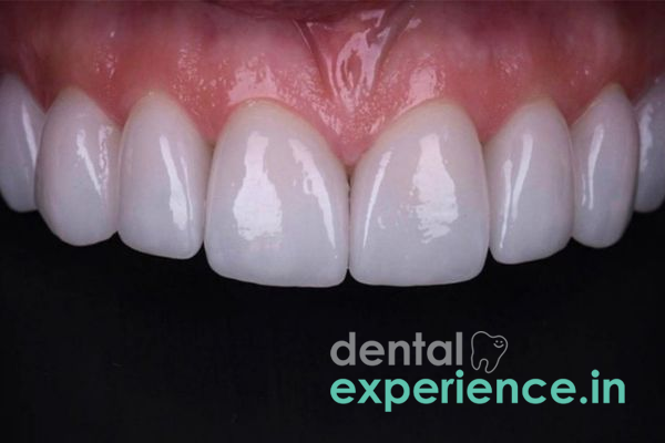
Moderate Damage - Treatment: Ceramic/Porcelain Veneers
Severe surface loss - Treatment: Monolithic Zirconia Crowns / Full Mouth Rehabilitation
Generally speaking, a complete full mouth reconstruction in Kerala will vary between Rs.250000/- to Rs.6000000/- based upon the extent of the case requirements and type of materials selected. High grade composite bonding options will generally cost between Rs.1500 -Rs.3500 per tooth, while superior durability options like e-max ceramic veneers or high strength monolithic zirconia crowns will generally cost between Rs.10000 - Rs.22000 per tooth.
Comprehensive treatment for widespread dental wear requires multi-disciplinary expertise. Patients are best served by a Prosthodontist who specializes in reconstructive dentistry and restoring functionality of the entire dentition working with a Cosmetic Dentist specializing in smile design and porcelain veneers.
Endodontists manage Root Canal Therapy for patients whose nerves were exposed due to severe dental wear. An Oral & Maxillo Facial Surgeon may assist in managing complicated cases that require joint realignment or structural modification to bone.
While enamel erosion develops slowly over years, delaying treatment can make subsequent intervention much more difficult. Once dentine is exposed, structural breakdown increases rapidly since dentine is much softer than enamel.
Delays in Treatment can result in nerve exposure; root canal infection; spontaneous fracture of the tooth, and tmj disorders that produce chronic pain. Early intervention reduces complexity and makes it possible for clinicians to utilize conservative bonding procedures instead of more complex full-mouth crown restorations.
Will damaged enamel regrow naturally?
No. Unlike skin or muscle tissue, damaged enamel does not contain living cells and therefore cannot regenerate without external assistance.
Do repeated episodes of teeth grinding at night result in permanent loss?
Yes. Repeated episodes of nocturnal clenching/grinding exert extremely high forces that flatten chewing surfaces over time.
What is the expected life-span for cosmetic restorative procedures?
Typically composite resins last 5-7 years while high-end ceramic/porcelain crowns/veneers last 15-20 years provided they receive regular maintenance care.
To manage severe tooth wear, one needs to go beyond cosmetics to restore proper functional relationships between components. We concentrate on establishing proper vertical occlusal relationships using highly biocompatible, durable monolithic zirconia restorations in order to prevent further deterioration and aesthetic results. Early detection facilitates utilization of conservative non-invasive approaches.














.jpg)
.jpg)

.jpg)
.jpg)

.jpg)
.jpg)
.jpg)
.jpg)
.jpg)
.jpg)
.jpg)
.jpg)