
 Treatment
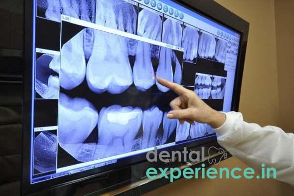
During a digital dental X-ray, a small sensor is placed inside your mouth to capture images of your teeth and jaw. These images are transmitted instantly to a computer, allowing your dentist to view and assess them promptly. This immediate feedback facilitates timely diagnosis and treatment planning.
Different imaging modalities are chosen based on clinical needs and complexity of the dental condition.
Patients who have undergone digital X-ray imaging will usually be able to get back to their daily day to day routines in an instant because the procedure is non-intrusive and causes little radiation exposure. Complications are rare, but if you experience any discomfort or have concerns, it is best to discuss them with your dental specialist. It is also important to note that follow-up visits are necessary to review the output of that X-ray and prepare for any other treatments moving forward.
Digital X-rays play an important role as one of the few innovative tools that seem to have truly transformed modern dentistry, providing a fast, safe and highly detailed form of dental imaging. The advent of digital sensors has made it possible for dental professionals in India and globally to improve the quality of images and minimize exposure to radiation by replacing traditional film-based X-rays. This technology is used for diagnostics, treatment planning, and to assist in patient care, representing a substantial advancement in dental radiography and a fundamental component of modern-day dental treatment.
Digital X-rays serve the main function of capturing clear and precise images of dental structures, such as teeth, bones, and surrounding soft tissues. These images are crucial for early diagnosis for cavities, bone resorption, infections and other dental problems. Enhanced diagnostic precision enables better treatment strategies and follow-ups, promoting the successful management of not only overall dental wellness, but more complex treatment protocols as well.
The digital x-ray technique is appropriate for a wide range of patients, from young to old. They are particularly beneficial for patients who need frequent imaging, such as
Digital radiography delivers the kind of clarity that helps doctors to accurately diagnosis and plan a treatment.
There are two main types of digital X-rays:
They are designed for different diagnostic requirements and ensure the appropriate imaging technique is employed with respect to the clinical scenario.
Digital X-ray acquisition is simple and patient friendly.
Digital radiography has many advantages over traditional radiography.
Digital X-ray equipment designed to meet the needs of a busy dental clinic. With proper maintenance and calibration, high-quality sensors and sophisticated imaging software are engineered for long-term performance. While technological advancements do happen here and there, well-maintained digital X-ray systems prove to be consistently reliable to the very end; thus, investing in a robust digital X-ray system is worth the spend for long for your dental practice.
Digital X-rays offer an impressive range of benefits, but one of the most remarkable is the swift speed of image capture and processing. Usually, the entire process takes only a few minutes. Intraoral digital X-rays can often be taken in less than a minute per exposure, and extraoral panoramic images can be captured quickly. Such efficiency not only improves patient comfort by limiting chair time but also enables dental professionals to swiftly review images and proceed with treatment planning.
Digital X-rays deliver clinical benefits as well as a cost-effective solution in the long run. Digital X-ray machines can be expensive with the initial investment lying between INR 2,00,000 to INR 5,00,000 depending on the brand and other features. The price of a digital X-ray test is affordable for patients in India. The cost of an intraoral digital x-ray is usually between INR 150 and INR 500 per exposure and extraoral or panoramic digital x-rays, INR 300-1500.
One of the most common radiographic modalities in dentistry are the digital X-rays. Digital X-ray machines, which emit much less radiation than regular film X-rays, making them safe even for sensitive groups such as children and pregnant women. As is the case with other radiographic procedures, repeated exposures should be tracked, but following strict safety protocols means that the potential risks remain exceptionally low.
Importantly, there are few limitations to digital X-ray technology, despite its many advantages.














.jpg)
.jpg)

.jpg)
.jpg)

.jpg)
.jpg)
.jpg)
.jpg)
.jpg)
.jpg)
.jpg)
.jpg)

